. 2018 Apr;41(4):1931-1938.
doi: 10.3892/ijmm.2018.3434. Epub 2018 Jan 29.

Extracorporeal shock wave therapy with low-energy flux density inhibits hypertrophic scar formation in an animal model

Affiliations

Extracorporeal shock wave therapy with low-energy flux density inhibits hypertrophic scar formation in an animal model

Jing-Chun Zhao et al. Int J Mol Med. 2018 Apr.

Abstract

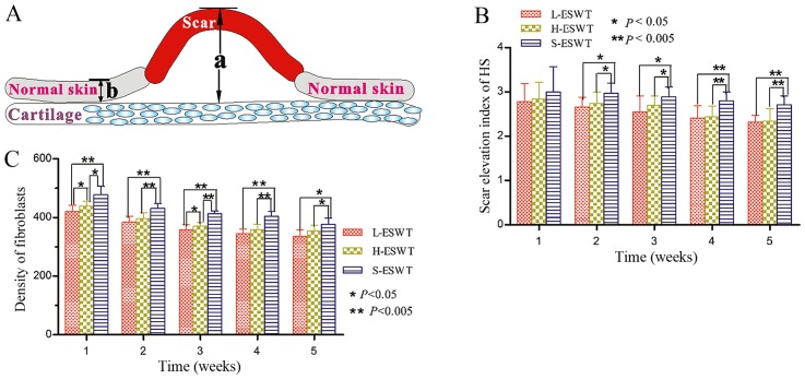
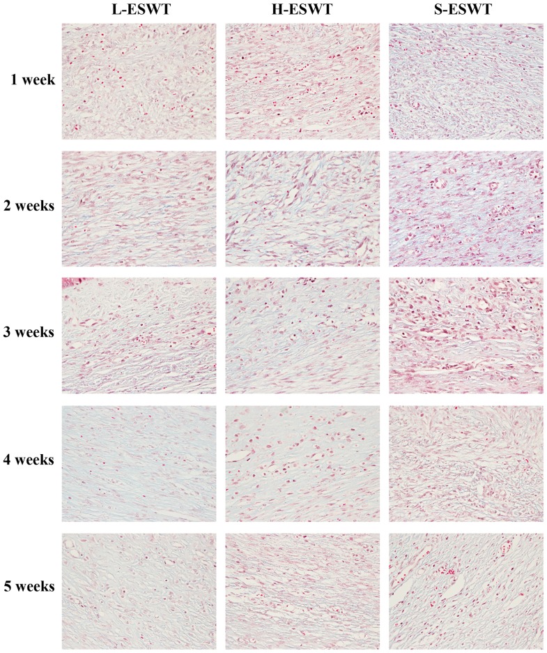
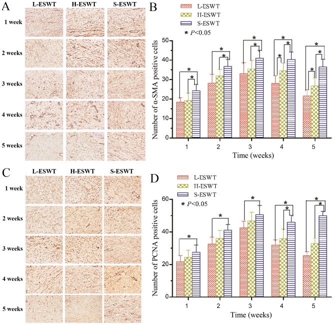
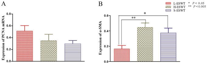
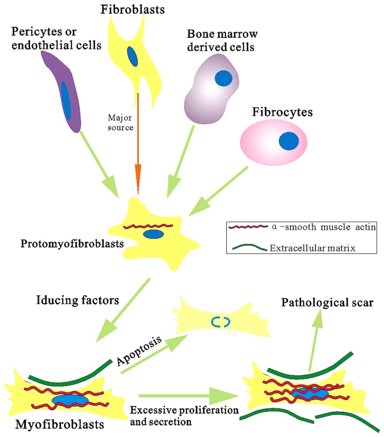
Hypertrophic scar is characterized by excessive deposits of collagen during skin wound healing, which could become a challenge to clinicians. This study assessed the effects of the extracorporeal shock wave therapy (ESWT) on hypertrophic scar formation and the underlying gene regu-lation. A rabbit ear hypertrophic scar model was generated and randomly divided into three groups: L-ESWT group to receive L-ESWT (energy flux density of 0.1 mJ/mm2), H-ESWT (energy flux density of 0.2 mJ/mm2) and sham ESWT group (S-ESWT). Hypertrophic scar tissues were then collected and stained with hematoxylin and eosin (H&E) and Masson's trichrome staining, respectively, to assess scar elevation index (SEI), fibroblast density and collagen fiber arrangement. Expression of cell proliferation marker proliferating cell nuclear antigen (PCNA) and α-smooth muscle actin (α-SMA) were assessed using RT-PCR and immunohistochemistry in hypertrophic scar tissues. H&E staining sections showed significant reduction of SEI and fibroblast density in both ESWT treatment groups compared to S-ESWT, but there was no dramatic difference between L-ESWT and H-ESWT groups. Masson's trichrome staining showed that collagen fibers were more slender and broader and oriented in parallel to skin surface after administration of ESWT compared to control tissues. At the gene level, PCNA‑positive fibroblasts and α-SMA-positive myofibroblasts were significantly decreased after L-ESWT or H-ESWT compared to the controls. Furthermore, there was no significant difference in expression of PCNA mRNA between L-ESWT or H-ESWT and S-ESWT, whereas expression of α-SMA mRNA significantly decreased in L-ESWT compared to that of H-ESWT and S-ESWT (P=0.002 and P=0.030, respectively). In conclusion, L-ESWT could be effective on suppression of hypertrophic scar formation by inhibition of scar elevation index and fibroblast density as well as α-SMA expression in hypertrophic scar tissues of the rabbit model.


Conflict of interest statement

Competing interests

The authors declare that they have no competing interests.

Figures

Figure 1
Figure 1
The rabbit ear model of hypertrophic scar and effects of extracorporeal shock wave therapy (ESWT). (A) The model of scar elevation index (SEI). a, The height from the peak of hypertrophic scar to the cartilage; b, the height from the surface of normal skin to the cartilage. SEI=a/b. (B) Effects of ESWT administration on SEI reduction in the rabbit model of hypertrophic scar. (C) Effects of ESWT administration on inhibition of fibroblasts density.
Figure 2
Figure 2
Effects of extracorporeal shock wave therapy (ESWT) administration on modulation of collagen arrangement in hypertrophic scar tissues. The animals were subjected to L-ESWT or H-ESWT for up to 5 weeks and the hypertrophic scar tissues were processed for Masson's trichrome staining and evaluation of collagen arrangement. Magnification, ×200.
Figure 3
Figure 3
Effects of extracorporeal shock wave therapy (ESWT) administration on suppression of proliferating cell nuclear antigen (PCNA) and α-smooth muscle actin (α-SMA) protein expression. The animals were subjected to L-ESWT or H-ESWT for up to 5 weeks and the hypertrophic scar tissues were processed for immunohistochemical analysis. (A and B) α-SMA and (C and D) PCNA. Magnification, ×200. The graphs are summarized data of the immunostaining.
Figure 4
Figure 4
Effects of extracorporeal shock wave therapy (ESWT) administration on modulation of proliferating cell nuclear antigen (PCNA) and α-smooth muscle actin (α-SMA) mRNA level. The animals were subjected to L-ESWT or H-ESWT for up to 5 weeks and the hypertrophic scar tissues were processed for RT-PCR analysis of (A) PCNA and (B) α-SMA mRNA.
Figure 5
Figure 5
Schematic diagram of myofibroblast sources and differentiation. Inducing factors, e.g., transforming growth factor-β (TGF-β), mechanical stress and integrin would induce fibroblasts change into protomyofibroblasts and then transition into differentiated myofibroblasts characterized by contractile properties because of the formation of robust stress fibers containing α-smooth muscle actin (α-SMA).

References

    1. Stadelmann WK, Digenis AG, Tobin GR. Physiology and healing dynamics of chronic cutaneous wounds. Am J Surg. 1998;176(Suppl 2A):26S–38S. doi: 10.1016/S0002-9610(98)00183-4. - DOI - PubMed
    1. Midwood KS, Williams LV, Schwarzbauer JE. Tissue repair and the dynamics of the extracellular matrix. Int J Biochem Cell Biol. 2004;36:1031–1037. doi: 10.1016/j.biocel.2003.12.003. - DOI - PubMed
    1. Gauglitz GG, Korting HC, Pavicic T, Ruzicka T, Jeschke MG. Hypertrophic scarring and keloids: pathomechanisms and current and emerging treatment strategies. Mol Med. 2011;17:113–125. doi: 10.2119/molmed.2009.00153. - DOI - PMC - PubMed
    1. Nedelec B, Shankowsky H, Scott PG, Ghahary A, Tredget EE. Myofibroblasts and apoptosis in human hypertrophic scars: the effect of interferon-alpha2b. Surgery. 2001;130:798–808. doi: 10.1067/msy.2001.116453. - DOI - PubMed
    1. Slemp AE, Kirschner RE. Keloids and scars: a review of keloids and scars, their pathogenesis, risk factors, and management. Curr Opin Pediatr. 2006;18:396–402. doi: 10.1097/01.mop.0000236389.41462.ef. - DOI - PubMed

MeSH terms