Review
. 2018 Feb 2;19(2):446.
doi: 10.3390/ijms19020446.

Temporomandibular Joint Regenerative Medicine

Affiliations
Review

Temporomandibular Joint Regenerative Medicine

Xavier Van Bellinghen et al. Int J Mol Sci. .

Abstract

The temporomandibular joint (TMJ) is an articulation formed between the temporal bone and the mandibular condyle which is commonly affected. These affections are often so painful during fundamental oral activities that patients have lower quality of life. Limitations of therapeutics for severe TMJ diseases have led to increased interest in regenerative strategies combining stem cells, implantable scaffolds and well-targeting bioactive molecules. To succeed in functional and structural regeneration of TMJ is very challenging. Innovative strategies and biomaterials are absolutely crucial because TMJ can be considered as one of the most difficult tissues to regenerate due to its limited healing capacity, its unique histological and structural properties and the necessity for long-term prevention of its ossified or fibrous adhesions. The ideal approach for TMJ regeneration is a unique scaffold functionalized with an osteochondral molecular gradient containing a single stem cell population able to undergo osteogenic and chondrogenic differentiation such as BMSCs, ADSCs or DPSCs. The key for this complex regeneration is the functionalization with active molecules such as IGF-1, TGF-β1 or bFGF. This regeneration can be optimized by nano/micro-assisted functionalization and by spatiotemporal drug delivery systems orchestrating the 3D formation of TMJ tissues.

Keywords: drug delivery systems; functionalization; growth factors; nanotechnology; osteochondral regeneration; regenerative medicine; scaffolds; stem cells; temporomandibular joint.


Conflict of interest statement

The authors declare no conflict of interest.

Figures

Figure 1
Figure 1
Invasive treatment of a patient suffering from TMJ (temporomandibular joint) ankylosis: Right lateral view of a 3D CT scan reconstruction of the head: the right TMJ is affected by joint space narrowing (A) and the left TMJ space has completely disappeared and been replaced by an osseous block (B). This replacement of the left TMJ by an osseous block of ankylosis is seen on transversal (C), and on coronal (D) CT scan sections. Intraoperative view of invasive treatment: the osseous bloc of ankylosis replacing the left TMJ space is approached through a pre-auricular incision (E). Preoperative intraoral photograph showing the absence of mouth opening (F).
Figure 2
Figure 2
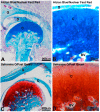
Histological organization of murine TMJ. TMJ is an articulation between the glenoid fossa of temporal bone (G) and mandibular condyle (C). TMJ disc cushioning articular mechanical stresses is fibrocartilaginous (D). TMJ condyle is made of a specific articular cartilage and a underlying bone containing blood vessels in medullar spaces (BV). Alcian Blue/Nuclear Fast Red specifically staining of mucopolysaccharides in blue (A,B) and Safranine O/Fast Green staining of cartilaginous proteoglycans in orange/red (C,D) highlight the osteochondral interface.
Figure 3
Figure 3
Scheme of the composition of the five compartments of TMJ to regenerate. Their cellular and macromolecular compositions differ of lot. An osteochondral molecular gradient of functionalization able to orchestrate the 3D formation of different TMJ tissues is the key of its regeneration.
Figure 4
Figure 4
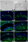
Expression of different conjunctive macromolecules in murine TMJ detected by immunofluorescence. TMJ observed by phase contrast microscope (A,B), Aggrecan expressed by chondrocytes in hypertrophic layer of mandibular condyle (C,D), type I collagen in the disc and in the fibrocartilage layer of mandibular condyle (E,F) and type II collagen in the fibrocartilage layer of mandibular condyle (G,H). Nuclei were stained with 4′,6-diamidino-2-phenylindole (DAPI). Condyle (C); Disc (D); Glenoid fossa (G).
Figure 5
Figure 5
Scanning electron microscopy (SEM) observations of nanofibrous pro-regenerative biomimetic implants: Poly(ε-caprolactone) implant with an electrospun nanofiber network mimicking the pattern of the connective tissue matrix (A); Poly(ε-caprolactone) implant functionalized with nanoreservoirs of growth factors on the surface of nanofibers (B). Scale bar: 3 µm.

References

    1. Gopal S.K., Shankar R., Vardhan B.H. Prevalence of temporo-mandibular disorders in symptomatic and asymptomatic patients: A cross-sectional study. Int. J. Adv. Health Sci. 2014;1:14–20.
    1. Su N., Liu Y., Yang X., Shen J., Wang H. Association of malocclusion, self-reported bruxism and chewing-side preference with oral health-related quality of life in patients with temporomandibular joint osteoarthritis. Int. Dent. J. 2017 doi: 10.1111/idj.12344. - DOI - PMC - PubMed
    1. Tanaka E., Detamore M.S., Mercuri L.G. Degenerative disorders of the temporomandibular joint: Etiology, diagnosis, and treatment. J. Dent. Res. 2008;87:296–307. doi: 10.1177/154405910808700406. - DOI - PubMed
    1. Aryaei A., Vapniarsky N., Hu J.C., Athanasiou K.A. Recent tissue engineering advances for the treatment of temporomandibular joint disorders. Curr. Osteoporos. Rep. 2016;14:269–279. doi: 10.1007/s11914-016-0327-y. - DOI - PMC - PubMed
    1. Zarb G.A., Carlsson G.E. Temporomandibular disorders: Osteoarthritis. J. Orofac. Pain. 1999;13:295–306. - PubMed

MeSH terms

Substances

LinkOut - more resources