. 2018 Feb 2;13(2):e0190245.
doi: 10.1371/journal.pone.0190245. eCollection 2018.

Regulating BMI1 expression via miRNAs promote Mesenchymal to Epithelial Transition (MET) and sensitizes breast cancer cell to chemotherapeutic drug

Affiliations

Regulating BMI1 expression via miRNAs promote Mesenchymal to Epithelial Transition (MET) and sensitizes breast cancer cell to chemotherapeutic drug

Nibedita Patel et al. PLoS One. .

Abstract

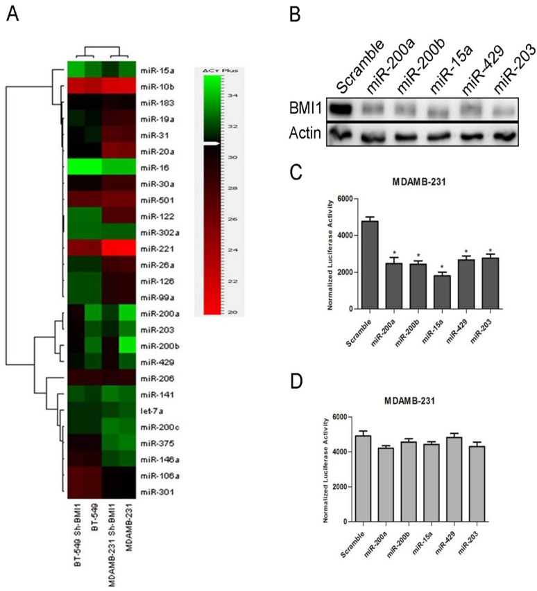
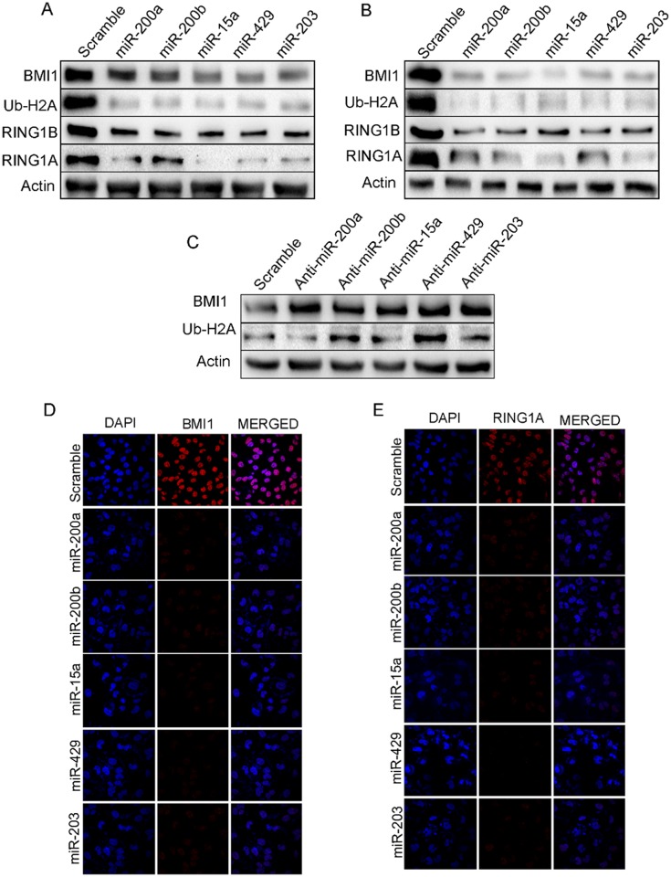
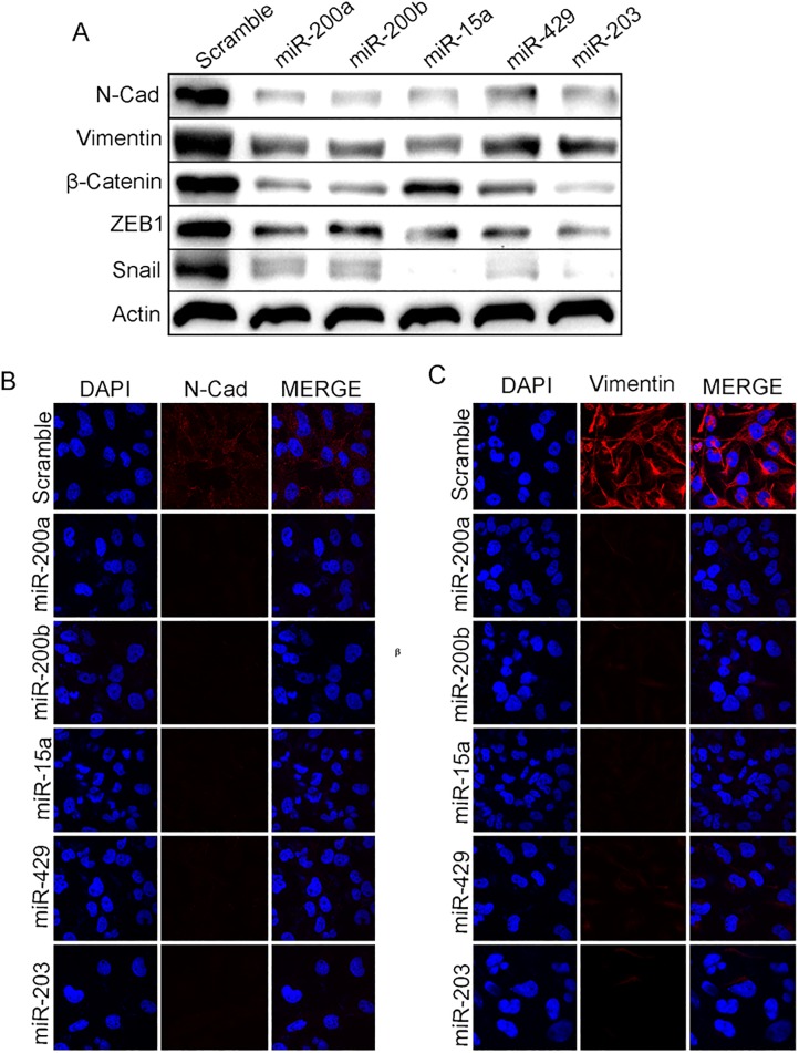
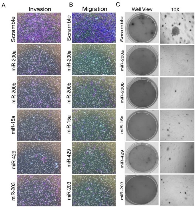
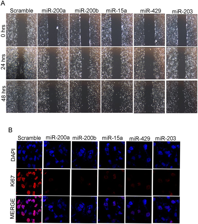
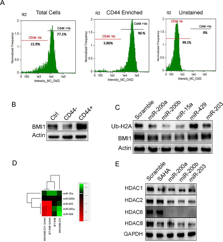
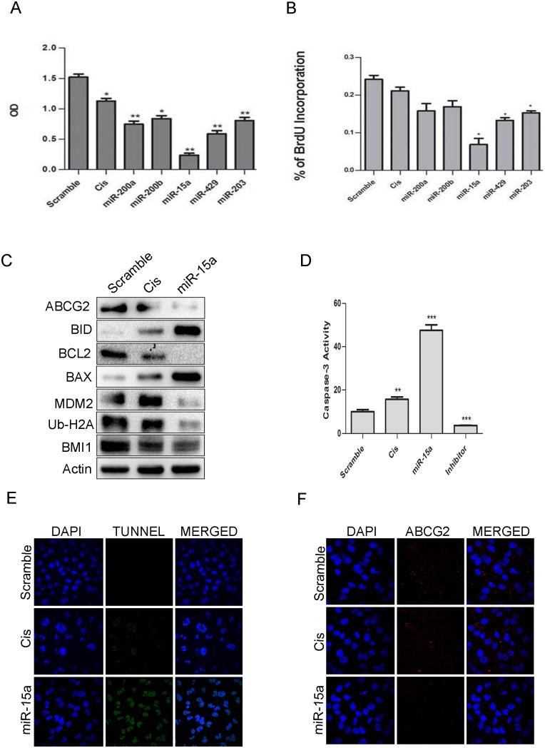
Polycomb group (PcG) proteinB lymphoma Mo-MLV insertion region 1 homolog (BMI1) is a transcriptional repressor that plays an important role in human carcinogenesis. MicroRNAs (miRNAs) are endogenous small non-coding RNAsthat implicate a negative regulation on gene expression. Deregulation of the expression of miRNAs has been implicated in tumorigenesis. Here, we have shown that knock-down ofBMI1increases theexpression of tumor-suppressivemiRNAs. Elevated levels of expression of miR-200a, miR-200b, miR-15a, miR-429, miR-203were observed upon knock-down of BMI1. Up-regulation of these miRNAsleads to down-regulation ofPRC1 group of proteins i.e. BMI1, RING1A, RING1B and Ub-H2A. Interestingly, overexpression of miR-200a, miR-200b and miR-15aalso produced decreased BMI1 and Ub-H2A protein expression in the CD44+ Cancer Stem Cellpopulation of MDAMB-231cells. Also,elevating the levels of BMI1 regulated miRNAspromoted Mesenchymal to Epithelial transition by regulating the expression of N-Cadherin, Vimentin, β-Catenin, Zeb, Snail thereby resulting in decreased invasion, migration and proliferation. Here, we also report that miR-200a, miR-200b, miR-203 accretes the sensitivity of MDAMB-231 cells to the histone deacetylase inhibitor (HDACi) SAHA and miR-15a sensitized breast cancer cells to the chemotherapeutic drug cisplatin leading to apoptosis. These findings suggest that modulatingspecific miRNAs may serve as a therapeutic approach for the treatment of breast cancer.


Conflict of interest statement

Competing Interests: The authors have declared that no competing interests exist.

Figures

Fig 1
Fig 1. BMI1 regulated by miRNAs.
Expression of the mentioned miRNAs in MDAMB-231 and BT-549 having pSMP-Bmi1 (A). Protein expression of BMI1wasanalyzed by performing western blotting in BT-549 cells having overexpressed miR-200a, miR-200b, miR-15a, miR-429, and miR-203. Actin was used as endogenous control (B).wt-BMI1 3’ UTR and Mut-BMI1 3’UTR were overexpressed along with miR-200a, miR-200b, miR-15a, miR-429, miR-203 and luciferase activity was measured (C,D).
Fig 2
Fig 2. miR-200a, miR-200b, miR-15a, miR-429, miR-203 regulate PRC1 proteins in MDAMB-231 cells.
Expression of BMI1, RING1A, RING1B and Ub-H2A in MDAMB-231(A) and BT549 (B) cells having overexpression of miR-200a, miR-200b, miR-15a, miR-429, miR-203. Expression of BMI1, Ub-H2A protein in MDAMB-231cells transfected with Anti-miR- 200a, Anti-miR-200b, Anti-miR-15a, Anti-miR-449, Anti-miR-203 (C) BMI1, RING1A localization in MDAMB-231 cells having overexpressed miR-200a, miR-200b, miR-15a, miR-449, miR-203under confocal microscopy (D, E). Cells transfected with Scramble Vector were used as controls. Bar indicates 200μm.
Fig 3
Fig 3. miR-200a, miR-200b, miR-429, and miR-203 induce Mesenchymal to Epithelial transition.
Level of expression of N-cadherin, Vimentin, β-Catenin, ZEB-1, Snailin MDAMB-231cells having overexpressed miR-200a, miR-200b, miR-15a, miR-429, and miR-203(A). Cytological localization of Vimentin, N-cadherin in MDAMB-231 cells having mentioned over expressed miRNAs.ScramblemiRNA vector was used as control (B, C).Bar indicates 200μm.
Fig 4
Fig 4. miRNAs inhibit migration, invasion and anchorage-dependent growth in MDAMB-231 cells.
Migratory and invasive rolesin MDAMB-231 cells having overexpressed miRNAs(A, B). Soft agar assay in MDAMB-231 cells having over expressed miRNAs (no of replicates 3) (C).Scramble miRNA vector transfected cells were used as control. All the experiments were performed in triplicates and standard deviation is represented in error bars.
Fig 5
Fig 5. miR-200a, miR-200b, miR-15a, miR-429, miR-203 inhibits migration and cell proliferation.
Images at 0hrs, 24 hrs and48 hrs of migration assay in MDAMB-231 cells having overexpressed miRNAs as well as transfected with scrambled miRNA vector control (A).Observation of Ki-67 localization under confocal microscopyin MDAMB-231 cellshaving over expressed miRNAs.ScramblemiRNA transfected cells were used as control (B).All the experiments were performed in triplicates.
Fig 6
Fig 6. miRNAs reduce expression of BMI1 and Ub-H2A in CD44+ stem cell population of MDAMB-231 cells and down-regulate HDACs expression in MDAMB-231 cells with SAHA treatment.
Segregation of CD44+ and CD44- cells by flow cytometry. From the CD44+ enriched population, 96% of the cells were CD44+ and 3.86% of the cells were CD44.Unstained cell population had 99.1% of CD44- cells (A). Expression of BMI1 in CD44+ and CD44- cells (B). Protein expression of BMI1 and Ub-H2A in CD44+ population transfected with miR-200a, miR-200b, miR-15a, miR-429 and miR-203 (C). Actin served as the loading control in (B) and (C). Levels of miR-200a, miR-200b, miR-15a, miR-429 and miR-203 in MDAMB-231 and BT-549 cells treated with SAHA. Untreated cells served as a control (D). Protein expression of various HDACs in cells having overexpression of miR-200a, miR-200b and miR-203. Cells with scramble miRNA were used as control. Cells without miRNA transfection but treated with SAHA served as positive control. GAPDH served as the loading control (E).
Fig 7
Fig 7. miR-15a sensitized MDAMB-231 cells to cisplatin.
Results of MTT assay showing the effect of miR-200a, miR-200b, miR-15a, miR-429, miR-203 in MDAMB-231 cells. Scramble miRNA vector was used as a control. Error bars indicate ±S.E of n = 3 (A). Results of BrdU assay showing the effect of miR-200a, miR-200b, miR-15a, miR-429, miR-203 in MDAMB-231 cells. Scramble miRNA vector was used as control. Error bars indicate ±S.E of n = 3 (B). Western blot analysis showing expression of BMI1, Ub-H2A, ABCG2, MDM2, pro-apoptotic and anti-apoptotic proteins like BAX, BID, BCL2, Caspase-3 in miR-15a overexpressed cells with cisplatin treatment. Scramble miRNA transfected cells served as control. β-Actin was used as gel loading control (C). Caspase-3 assay and Tunel assay were performed in MDAMB-231 containing ectopically expressed miR-15a with cisplatin treatment. Scramble miRNA transfected cells served as control. Cells with only cisplatin treatment served as positive control (D, E). Immunocytochemistry studies of above group of cells showing expression of ABCG2 (F).

References

    1. Pietersen AM, Evers B, Prasad AA, Tanger E, Cornelissen-Steijger P, Jonkers J, et al. Bmi1 regulates stem cells and proliferation and differentiation of committed cells in mammary epithelium. Current biology: CB. 2008;18(14):1094–9. Epub 2008/07/19. doi: 10.1016/j.cub.2008.06.070 . - DOI - PubMed
    1. Lukacs RU, Memarzadeh S, Wu H, Witte ON. Bmi-1 is a crucial regulator of prostate stem cell self-renewal and malignant transformation. Cell Stem Cell. 2010;7(6):682–93. Epub 2010/11/30. doi: 10.1016/j.stem.2010.11.013 - DOI - PMC - PubMed
    1. Ezhkova E, Pasolli HA, Parker JS, Stokes N, Su IH, Hannon G, et al. Ezh2 orchestrates gene expression for the stepwise differentiation of tissue-specific stem cells. Cell. 2009;136(6):1122–35. Epub 2009/03/24. doi: 10.1016/j.cell.2008.12.043 - DOI - PMC - PubMed
    1. Bracken AP, Pasini D, Capra M, Prosperini E, Colli E, Helin K. EZH2 is downstream of the pRB-E2F pathway, essential for proliferation and amplified in cancer. The EMBO journal. 2003;22(20):5323–35. Epub 2003/10/09. doi: 10.1093/emboj/cdg542 - DOI - PMC - PubMed
    1. Maynard MA, Ferretti R, Hilgendorf KI, Perret C, Whyte P, Lees JA. Bmi1 is required for tumorigenesis in a mouse model of intestinal cancer. Oncogene. 2014;33(28):3742–7. Epub 2013/08/21. doi: 10.1038/onc.2013.333 - DOI - PMC - PubMed

MeSH terms

Substances