. 2018 Feb 2;9(1):26.
doi: 10.1186/s13287-017-0763-3.

BCN057 induces intestinal stem cell repair and mitigates radiation-induced intestinal injury

Affiliations

BCN057 induces intestinal stem cell repair and mitigates radiation-induced intestinal injury

Payel Bhanja et al. Stem Cell Res Ther. .

Abstract

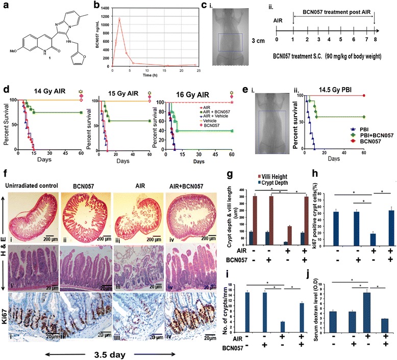
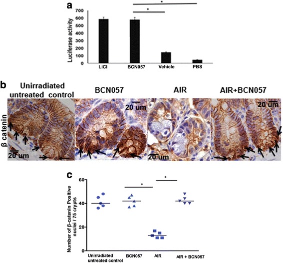
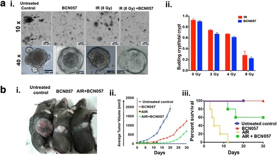
Background: Radiation-induced gastrointestinal syndrome (RIGS) results from the acute loss of intestinal stem cells (ISC), impaired epithelial regeneration, and subsequent loss of the mucosal barrier, resulting in electrolyte imbalance, diarrhea, weight loss, sepsis, and mortality. The high radiosensitivity of the intestinal epithelium limits effective radiotherapy against abdominal malignancies and limits the survival of victims of nuclear accidents or terrorism. Currently, there is no approved therapy to mitigate radiation toxicity in the intestine. Here we demonstrate that BCN057, an anti-neoplastic small molecular agent, induces ISC proliferation and promotes intestinal epithelial repair against radiation injury.

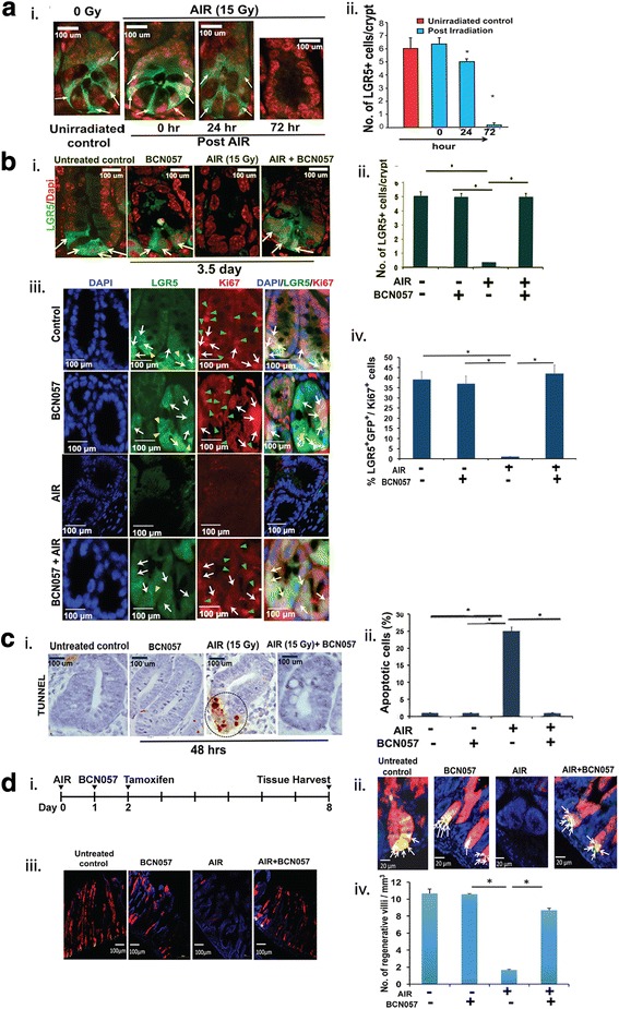
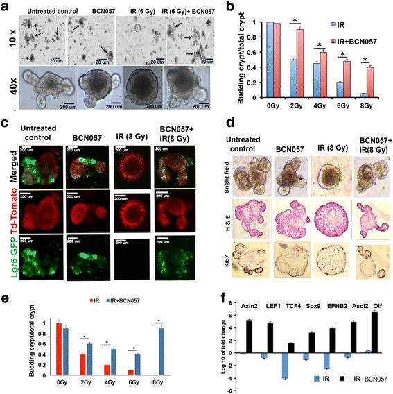
Methods: BCN057 (90 mg/kg body weight, subcutaneously) was injected into C57Bl6 male mice (JAX) at 24 h following abdominal irradiation (AIR) and was continued for 8 days post-irradiation. BCN057-mediated rescue of Lgr5-positive ISC was validated in Lgr5-EGFP-Cre-ERT2 mice exposed to AIR. The regenerative response of Lgr5-positive ISC was examined by lineage tracing assay using Lgr5-EGFP-ires-CreERT2-TdT mice with tamoxifen administration to activate Cre recombinase and thereby marking the ISC and their respective progeny. Ex vivo three-dimensional organoid cultures were developed from surgical specimens of human colon or from mice jejunum and were used to examine the radio-mitigating role of BCN057 on ISC ex vivo. Organoid growth was determined by quantifying the budding crypt/total crypt ratio. Statistical analysis was performed using Log-rank (Mantel-Cox) test and paired two-tail t test.

Results: Treatment with BCN057 24 h after a lethal dose of AIR rescues ISC, promotes regeneration of the intestinal epithelium, and thereby mitigates RIGS. Irradiated mice without BCN057 treatment suffered from RIGS, resulting in 100% mortality within 15 days post-radiation. Intestinal organoids developed from mice jejunum or human colon demonstrated a regenerative response with BCN057 treatment and mitigated radiation toxicity. However, BCN057 did not deliver radio-protection to mouse or human colon tumor tissue.

Conclusion: BCN057 is a potential mitigator against RIGS and may be useful for improving the therapeutic ratio of abdominal radiotherapy. This is the first report demonstrating that a small molecular agent mitigates radiation-induced intestinal injury by inducing ISC self-renewal and proliferation.

Keywords: Abdominal radiation; Intestinal stem cell; RIGS; Radiotherapy; Tumor.


Conflict of interest statement

Ethics approval

The present study is not considered as human subject research under HHS regulations at 45 CFR Part 46 and University of Kansas Human Subject Committee as we have used de-identified specimens and no living individual is involved as a study subject.

Consent for publication

Not applicable.

Competing interests

The authors declare that they have no competing interests.

Publisher’s Note

Springer Nature remains neutral with regard to jurisdictional claims in published maps and institutional affiliations.

Figures

Fig. 1
Fig. 1
BCN057 treatment at 24 h post-irradiation mitigates RIGS and improves survival in mice. a Chemical structure of BCN057 (3-[(Furan-2-ylmethyl)-amino]-2-(7-methoxy-2-oxo-1,2-dihydro-quinolin-3-yl)-6-methylimidazo[1,2-a]pyridin-1-ium). b Pharmacokinetics of a single injection of BCN057 90 mg/kg via s.c. administration in C57BL/6 mice. Cmax (obs) 1130.5 ng/mL, Tmax (obs) 2.0 h, Vss (expo) 15935.8 mL, CL (obs area) 700.075 mL/h. c Portal camera image demonstrating abdominal irradiation (AIR) exposure field (i) and BCN057 treatment schema (ii). A 3-cm area (indicated by the rectangular box) of the mouse containing the gastrointestinal tract was irradiated (irradiation field), while shielding the upper thorax, head, and neck, as well as the lower and upper extremities, and protecting a significant portion of the bone marrow, thus predominantly inducing radiation-induced gastrointestinal syndrome (RIGS). Mice exposed to AIR were treated with BCN057 (s.c.) (90 mg/kg body weight) at 24 h following irradiation and continued up to day 8 (single dose/day). d Kaplan-Meier survival analysis of C57BL/6 mice (n = 25/group) receiving vehicle, BCN057, or no treatment at 24 h after AIR (14 Gy, 15 Gy, or 16 Gy) and continued up to day 8. Mice receiving BCN057 after 14 Gy, 15 Gy, or 16 Gy AIR demonstrated 80%, 60%, and 40% survival, respectively, and they continued to survive beyond 60 days without any symptoms of gastroenteritis or any other health complications, whereas mice receiving vehicle or no treatment following AIR died within 15 days (p < 0.0003, p < 0.0004, and p < 0.0007, respectively, log-rank (Mantel-Cox) test). BCN057 or vehicle do not confer any toxicity to normal mice. e (i) Kaplan-Meier survival analysis of C57BL/6 mice (n = 25/group) exposed to partial body irradiation (PBI). Head, neck, and upper extremities were shielded to spare 40% of bone marrow (BM40%). The part of the body exposed to irradiation is indicated by a rectangular box. (ii) Mice receiving BCN057 at 24 h post-PBI demonstrated 70% survival compared with untreated controls (p < 0.0001 log-rank (Mantel-Cox) test). f H&E stained representative cross section of jejunum from C57BL/6 mice treated with BCN057 at 24 h post-AIR (upper panels). Note restitution of the epithelium in mice receiving BCN057 compared with irradiated controls. H&E stained representative transverse section of jejunum from C57BL/6 mice treated with BCN057 at 24 h post-AIR (middle panels). Note restitution of crypt villus structure in BCN057-treated mice. However, irradiated, untreated mice showed significant loss of crypts along with villi denudation. Representative Ki67 immunohistochemistry of mice jejunal sections (lower panels). Note the increase in Ki67-positive crypt cells in mice receiving BCN057 at 24 h after AIR (iv) compared with AIR controls (ii). g–i Histogram showing crypt depth and villi length (g), percentage of Ki67-positive crypt cells (h), and number of crypts per mm (i) in the jejunum. Irradiated mice receiving BCN057 at 24 h after AIR demonstrated an increase in crypt depth and villi length (*p < 0.0006), number of crypts (*p < 0.0004), and percentage of Ki67-positive crypt cells (*p < 0.0005) compared with irradiated controls. j Histogram demonstrating serum dextran level in C57BL/6 mice exposed to AIR and then treated with or without BCN057. Mice receiving BCN057 treatment demonstrated lower serum dextran levels, thereby suggesting restitution of epithelial integrity compared with irradiated untreated controls (*p < 0.004, n = 3 per group, unpaired t test, two-tailed). Unirradiated control mice and unirradiated BCN057 treated mice also showed lower serum dextran level compared with irradiated controls (*p < 0.006, *p < 0.005, unpaired t test, two-tailed)
Fig. 2
Fig. 2
BCN057 activates Wnt-β-catenin signaling. a HEK293 cells having TCF/LEF luciferase reporter construct were treated with BCN057 or LiCl. Treatment with BCN057 showed higher luciferase activity compared with vehicle control (*p < 0.001). b Representative microscopic images (×60 magnification) of jejunal sections immunostained with anti β-catenin antibody to determine β-catenin nuclear localization. Nuclei stained with hematoxylin. Irradiated mice treated with BCN057 demonstrated more nuclear β-catenin staining (dark brown; indicated with arrows) at the base of the crypt compared with control nuclei stained blue. c Nuclear β-catenin count. Each data point is the average of the number of β-catenin-positive nucleus from 15 crypts per field, five fields per mice. The number of β-catenin-positive nuclei in irradiated mice receiving BCN057 is higher compared with irradiated controls (*p < 0.005). Unirradiated mice receiving BCN057 showed a higher number of β-catenin-positive nuclei compared with irradiated controls (*p < 0.001; unpaired t test, two-tailed). AIR abdominal irradiation, PBS phosphate-buffered saline
Fig. 3
Fig. 3
BCN057 rescued Lgr5+ ISC and induced a regenerative response in vivo. a (i) Time-course study on the effect of abdominal irradiation (AIR) on Lgr5-positive ISC. Representative images of jejunal sections demonstrating the presence of green fluorescent protein (GFP)+Lgr5+ ISC (indicated with arrow) in Lgr5/GFP-IRES-Cre-ERT2 knock-in mice up to 24 h post-AIR. All the ISC at the crypt base disappeared at 72 h post-AIR. (ii) The number of Lgr5+GFP+ ISC per crypt in jejunal sections from Lgr5/GFP-IRES-Cre-ERT2 knock-in mice at different time points post-AIR. The number of Lgr5+GFP+ ISC per crypt reduced at 24 h post-irradiation (*p < 0.04). At 72 h post-AIR, most of the Lgr5+GFP+ ISC disappeared (p < 0.0001). b (i) Representative images of jejunal sections at 3.5 days post-AIR demonstrating the presence of GFP+Lgr5+ ISC (indicated with arrow) in Lgr5/GFP-IRES-Cre-ERT2 knock-in mice receiving BCN057 at 24 h post-AIR. Note the absence of GFP+ cells in mice receiving only AIR. (ii) The number of Lgr5+GFP+ ISC per crypt in jejunal sections from Lgr5/GFP-IRES-Cre-ERT2 knock-in mice exposed to irradiation and then treated with BCN057. The number of Lgr5+ cells are significantly higher in BCN057-treated irradiated mice compared with AIR controls (*p < 0.0001). Unirradiated mice receiving BCN057 also demonstrated a higher number of Lgr5+ cells at the crypt base compared with AIR controls (*p < 0.0002; unpaired t test, two-tailed). (iii) Representative images of jejunal sections demonstrating the presence of Ki67 in Lgr5+GFP+ ISC localized in the crypt base. Representative images from the single fluorescence channel showed localization of Lgr5+GFP+ cells (green, indicated with yellow arrow head) and Ki67+ cells (red, indicated with green arrow head). Cells that are double-positive for Ki67 and GFP are indicated with white arrows in both the single fluorescence channel and in the merged image. (iv) The percentage of Lgr5+GFP+/Ki67+ in jejunal sections from Lgr5/GFP-IRES-Cre-ERT2 knock-in mice exposed to irradiation and then treated with BCN057. The percentage of Lgr5+GFP+/Ki67+ cells are significantly higher in BCN057-treated irradiated mice compared with AIR controls (*p < 0.0001). Unirradiated mice receiving BCN057 also demonstrated a higher percentage of Lgr5+GFP+/Ki67+ cells at the crypt base compared with AIR controls (*p < 0.0003; unpaired t test, two-tailed). c (i) Representative image of jejunal sections at 48 h post-AIR demonstrating the presence of TUNEL-positive apoptotic cells at the crypt base in mice exposed to AIR. However, mice receiving the BCN057 treatment at 24 h post-AIR did not show any TUNEL-positive cells. (ii) Percentage of TUNEL-positive apoptotic cells in jejunal sections from mice exposed to AIR. The percentage of TUNEL-positive cells are significantly higher in the AIR group compared with mice receiving BCN057 at 24 h post-AIR (p < 0.0002). d (i) Schematic representation of the treatment schema for lineage tracing assay in Lgr5-eGFP-IRES-CreERT2; Rosa26-CAG-tdTomato mice. (ii) Confocal microscopic images (×40) of the jejunal section from Lgr5-eGFP-IRES-CreERT2; Rosa26-CAG-tdTomato mice. tdTomato (tdT)-positive cells are shown in red; Lgr5+GFP+ cells are shown in green. Nuclei are stained with DAPI (blue). Marked expansion of tdT-positive red cells above the +4 position (representing transit amplifying cells) were noted with BCN057 treatment. Please note the presence of yellow cells at the bottom of the crypt representing tdT-positive and GFP+Lgr5+ ISC (yellow due to red + green). (iii) Confocal microscopic images (×10) of the jejunal section from Lgr5-eGFP-IRES-CreERT2; Rosa26-CAG-tdTomato mice. Please note the presence of villi containing red tdT-positive cells (regenerative villi) in unirradiated controls or BCN057-treated mice. In the absence of BCN057 treatment, no tdT-positive cells were noted in irradiated mice jejunum. (iv) The number of regenerative villi. Irradiated mice receiving BCN057 showed a significantly higher number of regenerative villi compared with irradiated controls (*p < 0.002). Un-irradiated mice receiving BCN057 also demonstrated a higher number of regenerative villi compared with irradiated controls (*p < 0.0006)
Fig. 4
Fig. 4
BCN057 mitigates radiation toxicity in intestinal organoids derived from mice and human tissue. a Microscopic images (phase contrast) of intestinal organoids along with b a histogram of budding crypt/total crypt ratio demonstrating that BCN057 treatment improves organoid growth compared with irradiated (IR) controls (2 Gy, *p < 0.006; 4 Gy, *p < 0.008; 6 Gy, *p < 0.003; 8 Gy, *p < 0.002). Images at 10× (indicated with arrow) and 40× demonstrated the presence of a budding crypt in irradiated organoids treated with BCN057. c Confocal microscopic images of organoids developed from Lgr5-EGFP-CRE-ERT2; R26- ACTB-tdTomato-EGFP mice demonstrated that BCN057 treatment increases the presence of Lgr5-positive cells (green) in budding crypts compared with irradiated controls. tdTomato is constitutively expressed in these mice as membrane-bound protein, and therefore allows better visualization of cellular morphology. d Microscopic image (phase contrast) of organoids developed from human non-malignant colon demonstrating the loss of crypt domain exposed to irradiation (8 Gy). Both bright field and hematoxylin and eosin (H&E) staining demonstrated complete loss of budding crypt at 72 h post-irradiation. However, BCN057 treatment at 1 h post-radiation rescued the organoids from radiation toxicity and accelerated crypt-villus budding. Note the presence of a budding crypt-like structure in the BCN057-treated group. Ki67 staining demonstrated positive staining in the budding crypt-like structure in the BCN057-treated organoids indicating cell proliferation in this group. However, irradiated untreated organoids did not show any Ki67-positive budding crypt-like structure. e Ratio of budding crypts to total number of crypts in human colonic organoids is increased with BCN057 treatment in irradiated organoids compared with untreated irradiated controls (2 Gy, *p < 0.009; 4 Gy, *p < 0.006; 6 Gy, *p < 0.001; 8 Gy, *p < 0.003; unpaired t test, two-tailed). f qPCR analysis demonstrated that BCN057 treatment increases the mRNA levels of Wnt target genes in epithelial cells isolated from irradiated human colonic organoids. Data are the average of three human subjects
Fig. 5
Fig. 5
BCN057 does not have any radioprotective effect on colonic tumor tissue. a (i) BCN057 treatment did not rescue human malignant colonic organoids from radiation toxicity. Organoids derived from surgical specimens of colon tumor were exposed to irradiation (IR; 8 Gy) and then treated with BCN057. Note the loss of the budding crypt-like structure in irradiated organoids treated with BCN057. Treatment with BCN057 in unirradiated organoids also showed the loss of the budding crypt-like structure, indicating that BCN057 has an inhibitory effect on the growth and proliferation of malignant organoids. (ii) The effect of BCN057 treatment on the growth of irradiated crypt organoids developed from human colon tumor. Budding crypts to total crypt ratio reduced in a dose-dependent manner following irradiation (2–8 Gy). A similar pattern of budding crypts to total crypt ratio was observed in irradiated organoids with BCN057 treatment, indicating that BCN057 could not reduce the radiation toxicity in malignant colonic organoids. b BCN057 does not have radioprotective effect on mice abdominal tumors. (i) Representative image of C57BL/6 mice having MC38 colon tumor in the flank at day 20 post-abdominal irradiation (AIR). Note the reduction in tumor size in irradiated or unirradiated tumor treated with BCN057. Mice exposed to AIR without BCN057 treatment are not included in this image as they died within 12 days of AIR. (ii) Tumor growth curve demonstrating the effect of BCN057 treatment on mice abdominal MC38 tumors. Note the significant reduction in tumor growth in BCN057-treated mice following AIR compared with unirradiated untreated controls (p < 0.0004). BCN057 treatment in unirradiated mice also reduces the tumor growth compared with unirradiated untreated controls (p < 0.0007; n = 10/group). The tumor growth curve in the AIR group was discontinued as all mice died from radiation toxicity by day 12 post-radiation. (iii) Kaplan-Meier survival analysis of C57BL/6 mice (n = 10/group) with MC38 abdominal tumors receiving AIR, AIR + BCN057, BCN057, or no treatment. Please note that in the AIR control group all the mice died within 12 days post-radiation. Therefore, the tumor growth curve (ii) is not complete in these mice. Mice receiving BCN057 after 15 Gy AIR demonstrated 60% survival beyond day 20 post-irradiation (p < 0.0006; log-rank (Mantel-Cox) test). Untreated unirradiated control mice (blue line in the graph) were euthanized on day 20 as their tumor volume reached 2000 mm3 (the upper limit of tumor volume approved by the IACUC)

References

    1. Leibowitz BJ, Wei L, Zhang L, Ping X, Epperly M, Greenberger J, Cheng T, Yu J. Ionizing irradiation induces acute haematopoietic syndrome and gastrointestinal syndrome independently in mice. Nat Commun. 2014;5:3494. doi: 10.1038/ncomms4494. - DOI - PMC - PubMed
    1. Saha S, Bhanja P, Liu L, Alfieri AA, Yu D, Kandimalla ER, Agrawal S, Guha C. TLR9 agonist protects mice from radiation-induced gastrointestinal syndrome. PLoS ONE. 2012;7(1):e29357. doi: 10.1371/journal.pone.0029357. - DOI - PMC - PubMed
    1. Saha S, Aranda E, Hayakawa Y, Bhanja P, Atay S, Brodin NP, Li J, Asfaha S, Liu L, Tailor Y, et al. Macrophage-derived extracellular vesicle-packaged Wnts rescue intestinal stem cells and enhance survival after radiation injury. Nat Commun. 2016;7:13096. doi: 10.1038/ncomms13096. - DOI - PMC - PubMed
    1. Saha S, Bhanja P, Kabarriti R, Liu L, Alfieri AA, Guha C. Bone marrow stromal cell transplantation mitigates radiation-induced gastrointestinal syndrome in mice. PLoS ONE. 2011;6(9):e24072. doi: 10.1371/journal.pone.0024072. - DOI - PMC - PubMed
    1. Rios CI, Cassatt DR, Dicarlo AL, Macchiarini F, Ramakrishnan N, Norman MK, Maidment BW. Building the strategic national stockpile through the NIAID Radiation Nuclear Countermeasures Program. Drug Dev Res. 2014;75(1):23–28. doi: 10.1002/ddr.21163. - DOI - PMC - PubMed

Publication types

MeSH terms

Substances