. 2018 Feb 1;102(2):278-295.
doi: 10.1016/j.ajhg.2018.01.006.

OTUD7A Regulates Neurodevelopmental Phenotypes in the 15q13.3 Microdeletion Syndrome

Affiliations

OTUD7A Regulates Neurodevelopmental Phenotypes in the 15q13.3 Microdeletion Syndrome

Mohammed Uddin et al. Am J Hum Genet. .

Abstract

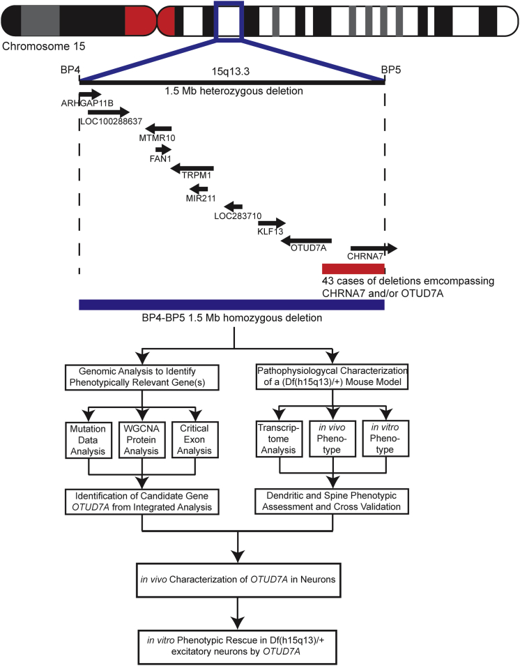
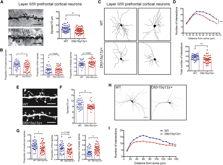
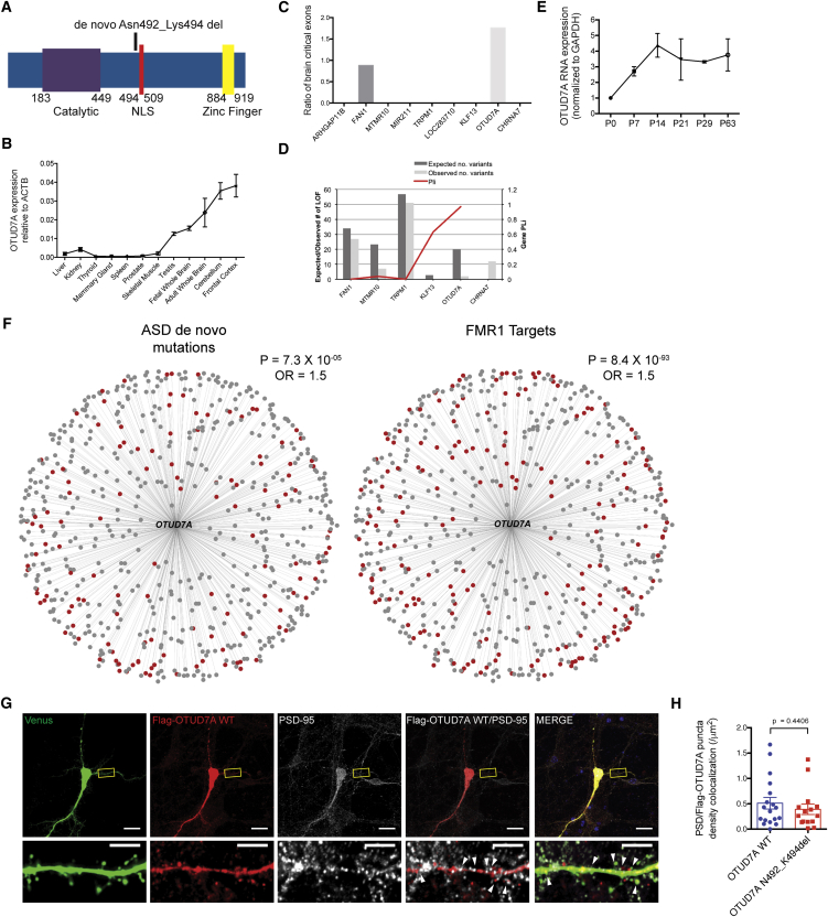
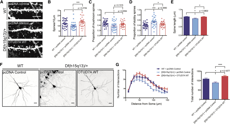
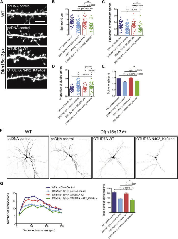
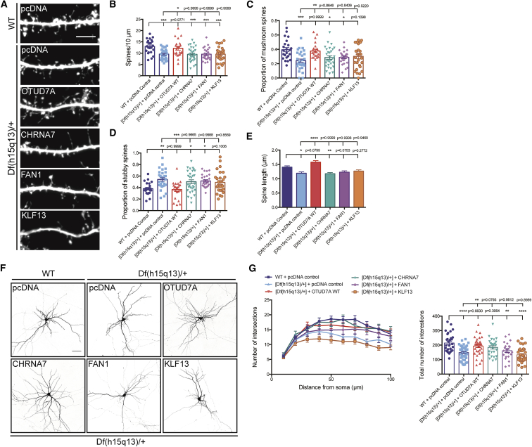
Copy-number variations (CNVs) are strong risk factors for neurodevelopmental and psychiatric disorders. The 15q13.3 microdeletion syndrome region contains up to ten genes and is associated with numerous conditions, including autism spectrum disorder (ASD), epilepsy, schizophrenia, and intellectual disability; however, the mechanisms underlying the pathogenesis of 15q13.3 microdeletion syndrome remain unknown. We combined whole-genome sequencing, human brain gene expression (proteome and transcriptome), and a mouse model with a syntenic heterozygous deletion (Df(h15q13)/+ mice) and determined that the microdeletion results in abnormal development of cortical dendritic spines and dendrite outgrowth. Analysis of large-scale genomic, transcriptomic, and proteomic data identified OTUD7A as a critical gene for brain function. OTUD7A was found to localize to dendritic and spine compartments in cortical neurons, and its reduced levels in Df(h15q13)/+ cortical neurons contributed to the dendritic spine and dendrite outgrowth deficits. Our results reveal OTUD7A as a major regulatory gene for 15q13.3 microdeletion syndrome phenotypes that contribute to the disease mechanism through abnormal cortical neuron morphological development.

Keywords: 15q13.3 microdeletion syndrome; OTUD7A; autism spectrum disorder; copy-number variation; dendrite; dendritic spine; deubiquitinase; neurodevelopmental disorder; schizophrenia; synapse.


Figures

Figure 1
Figure 1
Overview of the 15q13.3 Microdeletion and CHRNA7/OTUD7A Overlapping Deletion Schematic diagram showing the location of the human 15q13.3 locus and all ten genes within the 1.5 Mb BP4-BP5 deletion. Smaller deletions (red bar) are found within the BP4-BP5 region overlapping CHRNA7 and/or OTUD7A, modified from Lowther et al. The experimental workflow of the current study is presented below the schematic.
Figure 2
Figure 2
Df(h15q13)/+ Mice Have Defects in Dendritic Spine Development and Neuronal Morphology (A) Golgi-stained dendritic spine images (left) and spine density analysis (right). Df(h15q13)/+ mice show a decrease in spine density in layer II/III PFC neurons. WT, n = 40 dendritic segments, 22 neurons; Df(h15q13)/+, n = 40 dendritic segments, 26 neurons, 4 mice per condition, Student’s t test, ∗∗p < 0.01, t(78) = 2.846. Scale bar = 5 μm. (B) Df(h15q13)/+ mice show a decrease in PFC mushroom spines (left). WT, n = 40 dendritic segments, 22 neurons; Df(h15q13)/+, n = 40 dendritic segments, 26 neurons, 4 mice per condition, Student’s t test, p < 0.05, t(78) = 2.403, t(78) = 0.1312, t(78) = 1.673. (C) Traces of P28 WT and Df(h15q13)/+ Golgi-stained layer II/III prefrontal cortical (PFC) neurons. (D) Sholl analysis (left) and the total number of intersections (right). Df(h15q13)/+ mice show a decrease in dendrite growth in layer II/III PFC neurons. n = 40 neurons, 4 mice per condition, two-way ANOVA followed by Tukey’s post hoc test, ∗∗∗p < 0.001, F(9, 780) = 109.2, t(78) = 3.449. (E) Dendritic spine images from WT and Df(h15q13)/+ cultured neurons (scale bar = 5 μm). (F) Spine density measurements in cultured neurons. Spine density is decreased in Df(h15q13)/+ cultured cortical neurons. WT, n = 32 dendritic segments from 12 neurons; Df(h15q13)/+, n = 32 dendritic segments from 15 neurons, 3 cultures, Student’s t test, p < 0.05, t(62) = 2.311. (G) Dendritic spine classification in WT and Df(h15q13)/+ neurons. In Df(h15q13)/+ cultured cortical neurons, the proportion of mushroom-type spines is decreased (left) and the proportion of stubby type spines is increased (right). WT, n = 32 dendritic segments from 12 neurons; Df(h15q13)/+, n = 32 dendritic segments from 15 neurons, 3 cultures, Student’s t test, p < 0.05, ∗∗p < 0.01, t(62) = 2.007, t(62) = 0.1383, t(62) = 2.875. (H) DIV14 WT and Df(h15q13)/+ cultured cortical neurons expressing Venus (scale bar = 50 μm). (I) Sholl analysis of cultured cortical neurons. Dendrite growth is decreased in Df(h15q13)/+ neurons. n = 50 neurons, 3 cultures, two-way ANOVA followed by Sidak post hoc test, ∗∗∗p < 0.001, F(14, 1470) = 85.55. Error bars represent SEM.
Figure 3
Figure 3
Identification of OTUD7A as a Driver Gene of the 15q13.3 Microdeletion (A) Schematic of the human OTUD7A protein showing the exonic 9 bp mutation (de novo p.Asn492_Lys494 deletion in the protein) found in an ASD proband and affected sibling (proband is case 3 in Table 1). The mutation is located in the nuclear localization sequence (NLS) of OTUD7A. (B) OTUD7A mRNA expression relative to ACTB in various human tissues. OTUD7A is enriched in the brain, with highest expression in the frontal cortex. (C) Ratio of brain critical exons in nine genes within the 15q13.3 microdeletion region. FAN1 and OTUD7A contain brain critical exons, with OTUD7A showing a higher ratio than that of FAN1. (D) OTUD7A has the highest pLI (a score that indicates the probability that a gene is intolerant to a loss-of-function mutation) value compared to the other genes in the 15q13.3 critical region. The left y axis represents the number of observed LOF mutations within the ExAC population scale dataset and the right y axis shows the computed pLI score. (E) OTUD7A mRNA expression in the mouse brain is low in early postnatal life, increases into adolescence, and remains stable into adulthood (n = 2 technical replicates). (F) OTUD7A protein co-expression network module. Weighted gene correlation network analysis of OTUD7A from human proteome data shows that 616 genes are highly co-expressed with OTUD7A (top 25th percentile). 21% of genes highly co-expressed with OTUD7A harbor known ASD de novo mutations (left, red dots) and 30% of highly co-expressed genes are targets of FMR1 (right, red dots). (G) DIV14 WT mouse cortical neurons co-expressing Venus and FLAG-OTUD7A WT and co-stained for FLAG and PSD95. OTUD7A is co-localized with PSD95 in dendrites and dendritic spines. Arrows indicate co-localized puncta located in dendritic spines (scale bars = 20 μm, top; 5 μm, bottom). (H) Quantification of FLAG-OTUD7A and PSD95 puncta co-localization showed no changes between OTUD7A WT and OTUD7A p.Asn492_Lys494del overexpression (OTUD7A WT, n = 18 neurons; OTUD7A p.Asn492_Lys494del, n = 15 neurons, 2 cultures, Student’s t test, t(31) = 0.7813). Error bars represent SEM.
Figure 4
Figure 4
Reduced Expression of OTUD7A Contributes to Spine and Dendrite Defects in Df(h15q13)/+ Mice (A) Representative images of Venus-expressing dendritic spines from P22 WT or Df(h15q13)/+ neurons co-expressing Venus and pcDNA control or FLAG-OTUD7A WT (scale bar = 5 μm). (B) Expression of OTUD7A WT rescues dendrite spine density defects in Df(h15q13)/+ cortical neurons. WT + pcDNA control, n = 30 dendrite segments; [Df(h15q13)/+] + pcDNA, n = 30 dendrite segments; [Df(h15q13)/+] + OTUD7A WT, n = 30 dendrite segments; 3 cultures, one-way ANOVA followed by Tukey’s post hoc test, p < 0.05, ∗∗∗p < 0.001, F(2, 87) = 8.049. (C and D) Df(h15q13)/+ neurons show a decrease in mushroom spines and an increase of stubby spines compared to WT neurons. Expression of OTUD7A WT in Df(h15q13)/+ neurons decreases the proportion of stubby spines compared to Df(h15q13)/+ neurons. WT + pcDNA control, n = 30 dendrite segments; [Df(h15q13)/+] + pcDNA control, n = 30 dendrite segments; [Df(h15q13)/+] + OTUD7A WT, n = 30 dendrite segments; 3 cultures, one-way ANOVA followed by Tukey’s post hoc test, p < 0.05, ∗∗∗p < 0.001, F(2, 87) = 4.249, F(2, 87) = 8.636. (E) Df(h15q13)/+ neurons showed a decrease in spine length compared to WT neurons, and expression of OTUD7A WT in Df(h15q13)/+ neurons increased spine length. WT + pcDNA control, n = 586 spines; [Df(h15q13)/+] + pcDNA control, n = 475 spines; [Df(h15q13)/+] + OTUD7A WT, n = 664 spines; 3 cultures, one-way ANOVA followed by Tukey’s post hoc test, p < 0.05, ∗∗∗p < 0.001, F(2, 1722) = 3.956. (F) Representative images of littermate P22 WT and Df(h15q13)/+ neurons expressing Venus and pcDNA control or OTUD7A WT (scale bar = 50 μm). (G) Expression of OTUD7A WT rescues dendrite growth defects (number and total number of intersections) in Df(h15q13)/+ cortical neurons. WT + pcDNA control, n = 28 neurons, [Df(h15q13)/+] + pcDNA control, n = 24; [Df(h15q13)/+] + OTUD7A WT, n = 28 neurons; 3 cultures, one-way ANOVA followed by Tukey’s post hoc test, p < 0.05, ∗∗∗∗p < 0.0001, F(2, 61) = 13.53. Error bars represent SEM.
Figure 5
Figure 5
An Autism Spectrum Disorder-Linked De Novo Mutation in OTUD7A Disrupts Dendritic Spine Development and Neuronal Morphology (A) Dendritic spines from DIV14 WT and Df(h15q13)/+ cultured cortical neurons co-expressing Venus, and pcDNA control, FLAG-OTUD7A WT, or FLAG-OTUD7A p.Asn492_Lys494del (scale bar = 5 μm). (B) In Df(h15q13)/+ neurons, expression of OTUD7A p.Asn492_Lys494del does not rescue the reduction of dendritic spine density. WT + pcDNA, n = 32 dendritic segments, 18 neurons; pcDNA control, n = 48 dendritic segments, 30 neurons; OTUD7A WT, n = 38 dendritic segments, 21 neurons; OTUD7A p.Asn492_Lys494del, n = 28 dendritic segments, 19 neurons, 5 cultures, one-way ANOVA followed by Tukey’s post hoc test, ∗∗p < 0.01, ∗∗∗p < 0.001, F(3, 142) = 9.422. (C) Expression of OTUD7A p.Asn492_Lys494del does not increase the reduced proportion of mushroom spines in Df(h15q13)/+ neurons. n = same as (B), one-way ANOVA followed by Tukey’s post hoc test, ∗∗p < 0.01, ∗∗∗∗p < 0.0001, F(3, 141) = 11.99. (D) Expression of OTUD7A p.Asn492_Lys494del does not significantly decrease the increased proportion of stubby spines in Df(h15q13)/+ neurons. n = same as (B), one-way ANOVA followed by Tukey’s post hoc test, p < 0.05, ∗∗p < 0.01, F(3, 141) = 5.751. (E) Expression of OTUD7A p.Asn492_Lys494del is unable to increase the reduction in spine length in Df(h15q13)/+ neurons. WT + pcDNA, n = 653 spines; [Df(h15q13)/+] + pcDNA control, n = 770 spines; [Df(h15q13)/+] + OTUD7A WT, n = 712 spines; [Df(h15q13)/+] + OTUD7A p.Asn492_Lys393del, n = 365 spines, 5 cultures, one-way ANOVA followed by Tukey’s post hoc test, ∗∗p < 0.01, ∗∗∗∗p < 0.0001, F(3, 2496) = 13.20. (F) DIV14 WT and Df(h15q13)/+ cortical neurons expressing Venus and pcDNA control, FLAG-OTUD7A WT, or FLAG-OTUD7A p.Asn492_Lys494del (scale bar = 50 μm). (G) In Df(h15q13)/+ neurons, expression of OTUD7A p.Asn492_Lys494del does not rescue the reduction of dendrite growth. WT + pcDNA, n = 31 neurons; pcDNA control, n = 23 neurons; OTUD7A WT, n = 26 neurons; OTUD7A p.Asn492_Lys393del, n = 15 neurons, 5 cultures, one-way ANOVA followed by Tukey’s post hoc test, ∗∗p < 0.01, ∗∗∗p < 0.001, F(3, 133) = 9.613. Error bars represent SEM.
Figure 6
Figure 6
OTUD7A Is the Predominant Gene in the 15q13.3 Microdeletion Regulating Dendritic Spine Development (A) Dendritic spines from DIV14 WT and Df(h15q13)/+ cortical neurons co-expressing Venus and the indicated construct (scale bar = 5 μm). (B–E) Expression of CHRNA7, FAN1, or KLF13 in Df(h15q13)/+ neurons did not rescue the defects in certain phenotypes. (B) Dendritic spine density. WT + pcDNA control, n = 23 dendritic segments; [Df(h15q13)/+] + pcDNA control, n = 24 dendritic segments; [Df(h15q13)/+] + OTUD7A WT, n = 22 dendritic segments; [Df(h15q13)/+] + CHRNA7, n = 30 dendritic segments; [Df(h15q13)/+] + FAN1, n = 24 dendritic spines; [Df(h15q13)/+] + KLF13, n = 30 dendritic segments; 3 cultures, one-way ANOVA followed by Tukey’s post hoc test, p < 0.05, ∗∗p < 0.01, ∗∗∗p < 0.001, F(5, 157) = 6.907. (C and D) Proportion of mushroom spines (C) and the proportion of stubby spines (D). WT + pcDNA control, n = 23 dendritic segments; [Df(h15q13)/+] + pcDNA control, n = 24 dendritic segments; [Df(h15q13)/+] + OTUD7A WT, n = 22 dendritic segments; [Df(h15q13)/+] + CHRNA7, n = 30 dendritic segments; [Df(h15q13)/+] + FAN1, n = 24 dendritic spines; [Df(h15q13)/+] + KLF13, n = 30 dendritic segments; 3 cultures, one-way ANOVA followed by Tukey’s post hoc test, p < 0.05, ∗∗p < 0.01, F(5, 156) = 5.045, F(5, 157) = 5.666. (E) Spine length. WT + pcDNA control, n = 484 spines; [Df(h15q13)/+] + pcDNA control, n = 379 spines; [Df(h15q13)/+] + OTUD7A WT, n = 444 spines; [Df(h15q13)/+] + CHRNA7, n = 454 spines; [Df(h15q13)/+] + FAN1, n = 374 spines; [Df(h15q13)/+] + KLF13, n = 457 spines; 3 cultures, one-way ANOVA followed by Tukey’s post hoc test, p < 0.05, ∗∗p < 0.01, ∗∗∗∗p < 0.0001, F(5, 2728) = 10.64. (F) DIV14 WT and Df(h15q13)/+ cortical neurons co-expressing Venus and the indicated construct (scale bar = 50 μm). (G) Expression of OTUD7A WT or CHRNA7 in Df(h15q13)/+ cortical neurons rescues dendritic arborization defects, whereas FAN1 and KLF13 did not. WT + pcDNA, n = 30 neurons, [Df(h15q13)/+] + pcDNA control, n = 40 neurons; [Df(h15q13)/+] + OTUD7A WT, n = 35 neurons; [Df(h15q13)/+] + CHRNA7, n = 26 neurons; [Df(h15q13)/+] + FAN1, n = 23 neurons; [Df(h15q13)/+] + KLF13, n = 33 neurons, 3 cultures, one-way ANOVA followed by Tukey’s post hoc test, p < 0.05, F(5, 181) = 6.873. Error bars represent SEM.

References

    1. Uddin M., Pellecchia G., Thiruvahindrapuram B., D’Abate L., Merico D., Chan A., Zarrei M., Tammimies K., Walker S., Gazzellone M.J. Indexing effects of copy number variation on genes involved in developmental delay. Sci. Rep. 2016;6:28663. - PMC - PubMed
    1. Liu H., Abecasis G.R., Heath S.C., Knowles A., Demars S., Chen Y.J., Roos J.L., Rapoport J.L., Gogos J.A., Karayiorgou M. Genetic variation in the 22q11 locus and susceptibility to schizophrenia. Proc. Natl. Acad. Sci. USA. 2002;99:16859–16864. - PMC - PubMed
    1. Mukai J., Dhilla A., Drew L.J., Stark K.L., Cao L., MacDermott A.B., Karayiorgou M., Gogos J.A. Palmitoylation-dependent neurodevelopmental deficits in a mouse model of 22q11 microdeletion. Nat. Neurosci. 2008;11:1302–1310. - PMC - PubMed
    1. Portmann T., Yang M., Mao R., Panagiotakos G., Ellegood J., Dolen G., Bader P.L., Grueter B.A., Goold C., Fisher E. Behavioral abnormalities and circuit defects in the basal ganglia of a mouse model of 16p11.2 deletion syndrome. Cell Rep. 2014;7:1077–1092. - PMC - PubMed
    1. Blizinsky K.D., Diaz-Castro B., Forrest M.P., Schürmann B., Bach A.P., Martin-de-Saavedra M.D., Wang L., Csernansky J.G., Duan J., Penzes P. Reversal of dendritic phenotypes in 16p11.2 microduplication mouse model neurons by pharmacological targeting of a network hub. Proc. Natl. Acad. Sci. USA. 2016;113:8520–8525. - PMC - PubMed

Publication types

MeSH terms

Supplementary concepts

Grants and funding