. 2018 Feb 2;8(1):2267.
doi: 10.1038/s41598-018-20425-x.

PCSK9 induces a pro-inflammatory response in macrophages

Affiliations

PCSK9 induces a pro-inflammatory response in macrophages

Chiara Ricci et al. Sci Rep. .

Abstract

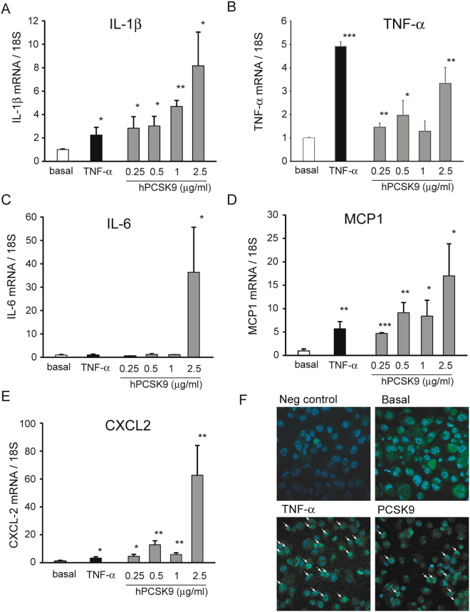
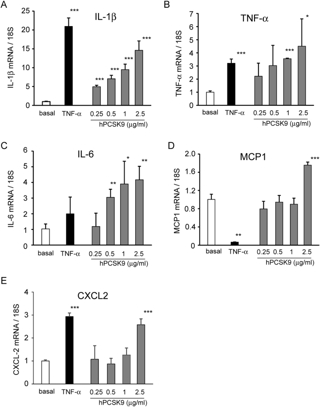
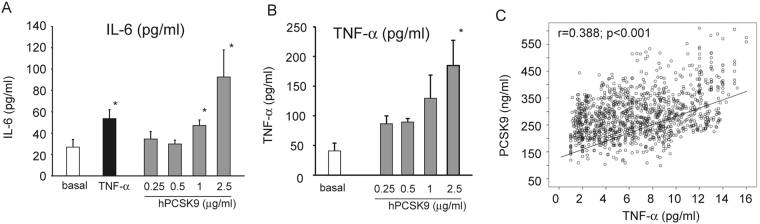
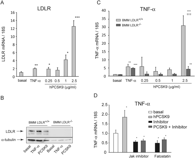
Intraplaque release of inflammatory cytokines from macrophages is implicated in atherogenesis by inducing the proliferation and migration of media smooth muscle cells (SMCs). PCSK9 is present and released by SMCs within the atherosclerotic plaque but its function is still unknown. In the present study, we tested the hypothesis that PCSK9 could elicit a pro-inflammatory effect on macrophages. THP-1-derived macrophages and human primary macrophages were exposed to different concentrations (0.250 ÷ 2.5 µg/ml) of human recombinant PCSK9 (hPCSK9). After 24 h incubation with 2.5 µg/ml PCSK9, a significant induction of IL-1β, IL-6, TNF-α, CXCL2, and MCP1 mRNA, were observed in both cell types. Co-culture of THP-1 macrophages with HepG2 overexpressing hPCSK9 also showed the induction of TNF-α (2.4 ± 0.5 fold) and IL-1β (8.6 ± 1.8 fold) mRNA in macrophages. The effect of hPCSK9 on TNF-α mRNA in murine LDLR-/- bone marrow macrophages (BMM) was significantly impaired as compared to wild-type BMM (4.3 ± 1.6 fold vs 31.1 ± 6.1 fold for LDLR-/- and LDLR+/+, respectively). Finally, a positive correlation between PCSK9 and TNF-α plasma levels of healthy adult subjects (males 533, females 537) was observed (B = 8.73, 95%CI 7.54 ÷ 9.93, p < 0.001). Taken together, the present study provides evidence of a pro-inflammatory action of PCSK9 on macrophages, mainly dependent by the LDLR.


Conflict of interest statement

The authors declare that they have no competing interests.

Figures

Figure 1
Figure 1
Recombinant hPCSK9 induces the expression of pro-inflammatory cytokines and chemokines in THP-1 derived macrophages. THP-1 macrophages were incubated for 24 h with TNF-α 10 ng/ml (as positive control) or different concentrations of recombinant hPCSK9 (0.25, 0.5, 1 and 2.5 μg/ml). At the end of the incubation, total RNA was extracted and (A) IL-1β, (B) TNF-α, (C) IL-6, (D) MCP-1 and (E) CXCL2 mRNA expression was determined by qRT-PCR. (F) THP-1 macrophages were incubated with TNF-α 10 ng/ml (as positive control) and recombinant hPCSK9 (2.5 μg/ml). After 24 h cells were fixed and immunostaining was performed for NF-κB p65 (blue: nuclei; green: p65 NF-κB). Arrows indicate cells with positive nuclear staining of p65. Data are given as mean ± SD of three independent experiments. Differences vs basal were assessed by Student’s t-test: *p < 0.05; **p < 0.01; ***p < 0.001.
Figure 2
Figure 2
Recombinant hPCSK9 induces the expression of pro-inflammatory cytokines and chemokines in human macrophages. Human macrophages were incubated for 24 h with TNF-α 10ng/ml (as positive control) or different concentrations of recombinant hPCSK9 (0.25, 0.5, 1 and 2.5 μg/ml). At the end of the incubation, total RNA was extracted and (A) IL-1β, (B) IL-6, (C) TNF-α, (D) MCP-1 and (E) CXCL2 mRNA expression was determined by qRT-PCR. Data are given as mean ± SD of three independent experiments. Differences vs basal were assessed by Student’s t-test: *p < 0.05; **p < 0.01; ***p < 0.001.
Figure 3
Figure 3
Effect of recombinant hPCSK9 on the release of IL-6 and TNF-α from human macrophages. Human monocytes were seeded in 6 well plates in RPMI containing 10% of the volunteer autologous serum and incubated for 1 week. Human macrophages were then incubated for 24 h with TNF-α 10 ng/ml (as positive control) or different concentrations of recombinant hPCSK9 (0.25, 0.5, 1 and 2.5 μg/ml). IL-6 (A) and TNF-α (B) protein amount was then assessed by ELISA assay on collected media. Results were expressed as pg of protein for ml of media. (C) Relationship between TNF-α and PCSK9 serum levels determined by ELISA assay from human samples. Data of panel A and B are given as mean ± SD of three independent experiments. Differences vs basal were assessed by Student’s t-test: *p < 0.05; **p < 0.01; ***p < 0.001.
Figure 4
Figure 4
PCSK9 released from HepG2 increased TNF-α and IL-1β gene expression in THP-1 derived macrophages. THP-1 macrophages were co-cultured with HepG2 or HepG2PCSK9 seeded on top of the transwell system. After 24 h, total RNA was extracted from THP-1 macrophages and TNF-α and IL-1β gene expression was evaluated by qRT-PCR. Data are given as mean ± SD of three independent experiments. Differences were assessed by Student’s t-test: *p < 0.05; **p < 0.01; ***p < 0.001.
Figure 5
Figure 5
Effect of recombinant hPCSK9 on TNF-α mRNA expression in LDLR−/− BMM. LDLR+/+ and LDLR−/− BMM were incubated with TNF-α 10 ng/ml (as positive control) or different concentrations of recombinant hPCSK9 (0.25, 0.5, 1 and 2.5 μg/ml). After 24 h, total RNA and protein extracts were prepared. LDLR receptor mRNA (A) and protein (B) expressions were evaluated by qRT-PCR and western blotting, respectively. (C) TNF-α mRNA levels were determined by qRT-PCR. (D) THP-1 macrophages were incubated with hPCSK9 (2.5 μg/ml) in the presence or absence of the JAK inhibitor (10 µM) and fatostatin (100 µM). Data are given as mean ± SD of three independent experiments. Differences vs basal (*) and between genotypes (†) were assessed by 1-way ANOVA: *p < 0.05; **p < 0.01; ***p < 0.001. p < 0.05; ††p < 0.01; †††p < 0.001.

References

    1. Williams KJ, Tabas I. The response-to-retention hypothesis of early atherogenesis. Arterioscler Thromb Vasc Biol. 1995;15:551–561. doi: 10.1161/01.ATV.15.5.551. - DOI - PMC - PubMed
    1. Hansson GK, Libby P, Tabas I. Inflammation and plaque vulnerability. Journal of internal medicine. 2015;278:483–493. doi: 10.1111/joim.12406. - DOI - PMC - PubMed
    1. Gordon S, Martinez FO. Alternative activation of macrophages: mechanism and functions. Immunity. 2010;32:593–604. doi: 10.1016/j.immuni.2010.05.007. - DOI - PubMed
    1. Yvan-Charvet L, et al. Increased inflammatory gene expression in ABC transporter-deficient macrophages: free cholesterol accumulation, increased signaling via toll-like receptors, and neutrophil infiltration of atherosclerotic lesions. Circulation. 2008;118:1837–1847. doi: 10.1161/CIRCULATIONAHA.108.793869. - DOI - PMC - PubMed
    1. Zhu X, et al. Increased cellular free cholesterol in macrophage-specific Abca1 knock-out mice enhances pro-inflammatory response of macrophages. J Biol Chem. 2008;283:22930–22941. doi: 10.1074/jbc.M801408200. - DOI - PMC - PubMed

Publication types