. 2018 Feb 2;8(1):2218.
doi: 10.1038/s41598-018-20672-y.

Focused shockwave induced blood-brain barrier opening and transfection

Affiliations

Focused shockwave induced blood-brain barrier opening and transfection

Yi Kung et al. Sci Rep. .

Abstract

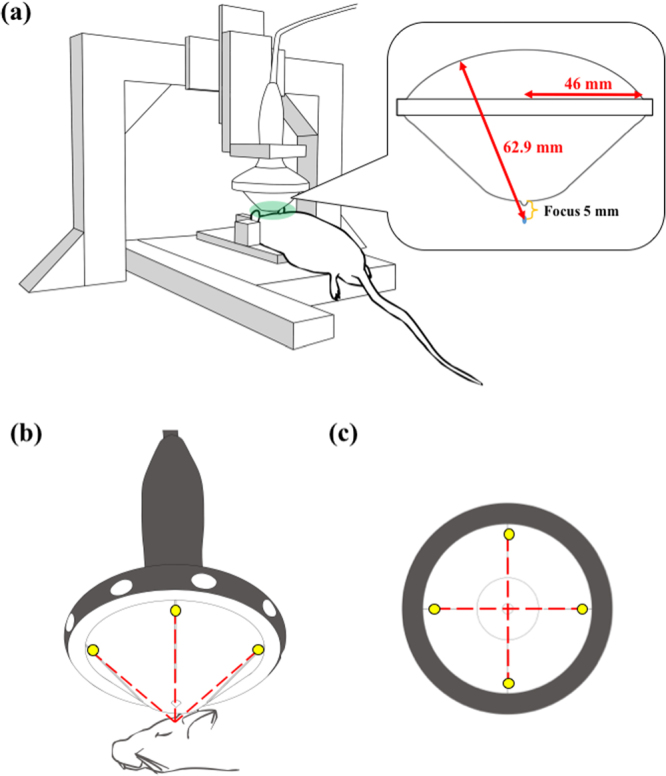
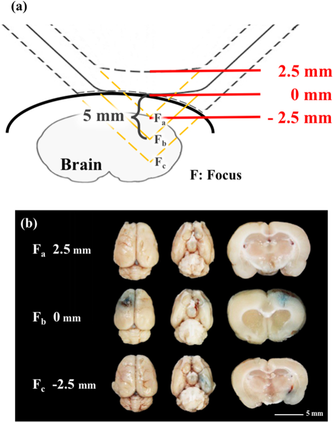
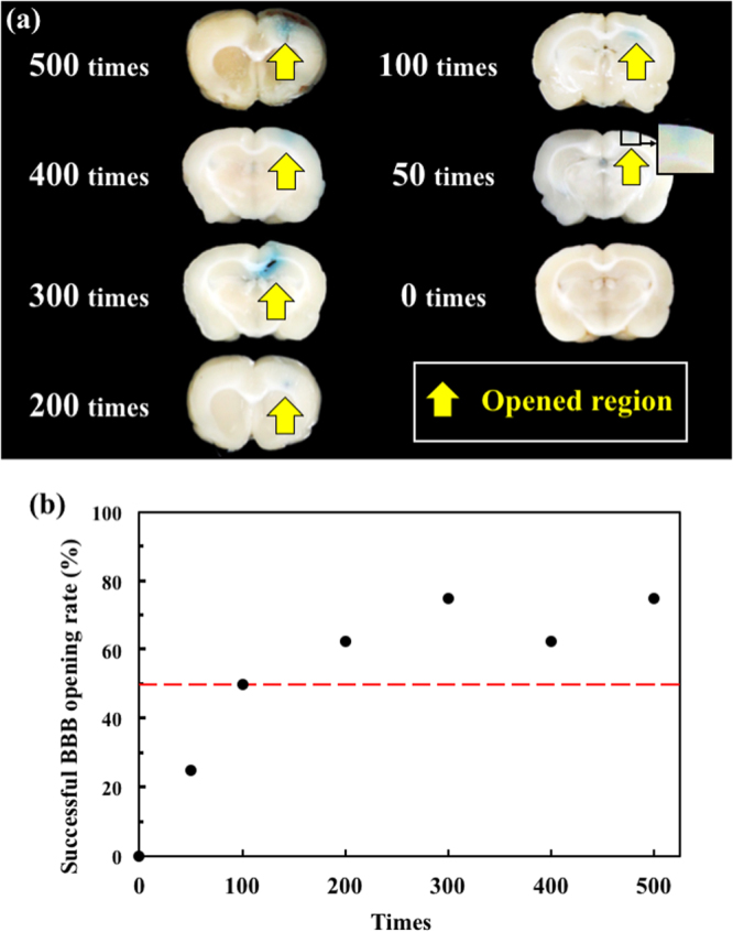
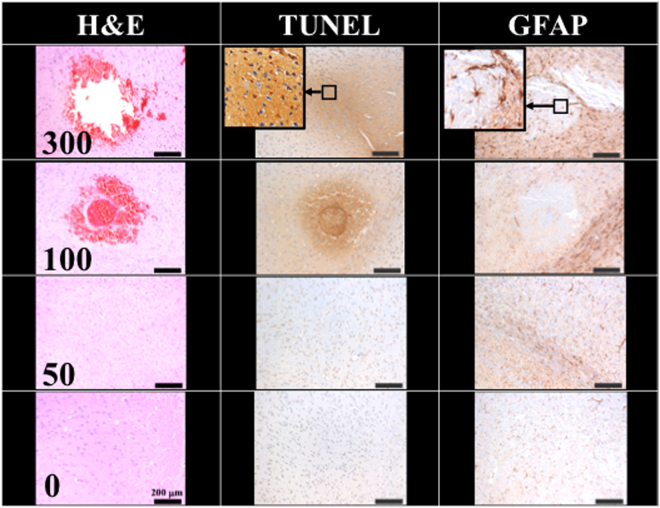
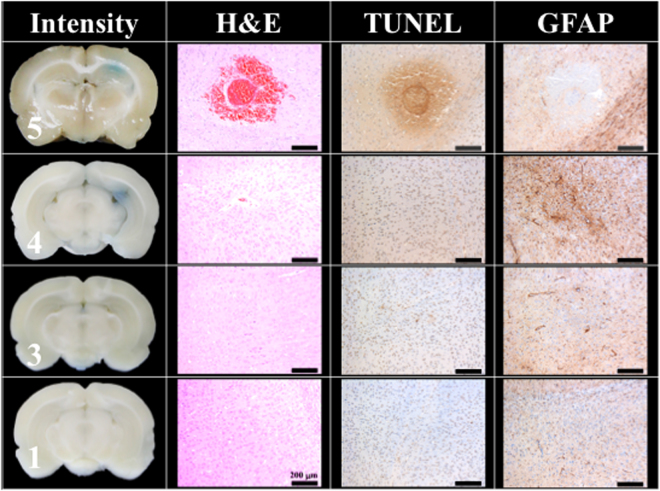
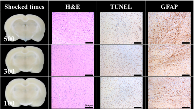
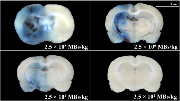
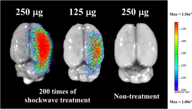
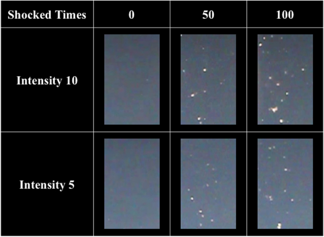
Despite extensive efforts in recent years, the blood-brain barrier (BBB) remains a significant obstacle for drug delivery. This study proposes using a clinical extracorporeal shockwave instrument to open the BBB, combined with a laser assisted bi-axial locating platform to achieve non-invasive, controllable-focus and reversible BBB opening in the brains of rats. Under shockwave treatment with an intensity level of 5 (P-9.79 MPa, energy flux density (EFD) 0.21 mJ/mm2) and a pulse repetition frequency of 5 Hz, the BBB could be opened after 50 shocks without the use of an ultrasound contrast agent. With the proposed method, the BBB opening can be precisely controlled in terms of depth, size and location. Moreover, a shockwave based gene transfection was demonstrated using a luciferase gene.


Conflict of interest statement

The authors declare that they have no competing interests.

Figures

Figure 1
Figure 1
(a) The shockwave probe positioning platform and its experimental implementation; (b) experimental implementation of the shockwave probe and the head of the rat, wherein the yellow dots are the positioning lasers and the red dotted lines are the lasers trajectories; (c) bottom view of the shockwave probe, in which all experiments were processed with a shockwave focus positioned 5 mm from the interface of the rat’s scalp and the bottom of the gel pad.
Figure 2
Figure 2
(a) Schematic of different penetration depths from the focus shockwave probe; (b) BBB opening at various distances from the scalp (2.5 mm, 0 mm, −2.5 mm). Shockwave treatment was carried out at an intensity level of 5 at PRF 5 Hz, in 500 iterations (N = 5). Scale bar was 5 mm.
Figure 3
Figure 3
(a) Histology sections at 0, 50, 100, 200, 300, 400, and 500 shockwave treatment iterations; (b) successful BBB opening rate under 0, 50, 100, 200, 300, 400, and 500 shockwave treatment iterations. Shockwave treatment was conducted with an intensity level of 5 and PRF of 5 Hz. N = 8.
Figure 4
Figure 4
Histology sections of H&E, TUNEL, and GFAP stain for 0, 50, 100, 300 shocks with an intensity level of 5 and PRF 5 Hz. Scale bar was 200 μm. N = 8. Dashed circle indicates a transverse cut of a vessel.
Figure 5
Figure 5
Histology sections of H&E, TUNEL, and GFAP stains for 100 shocks under intensity levels of 1, 3, 4, and 5. The PRF was set at 5 Hz. (Scale bar 200 μm, N = 5).
Figure 6
Figure 6
No BBB opening was found 24 hrs after 100, 300, 500 shocks at an intensity level of 5 and PRF 5 Hz. The indicator was 3% of Evans blue (pre-dissolved in 0.9% saline). Scale bar was 200 μm. N = 5.
Figure 7
Figure 7
Histology sections after 50 shocks with different concentrations of UCA (SonoVue). Shockwaves were administered at an intensity level of 5, PRF 5 Hz. The indicator was 3% of Evans blue (pre-dissolved in 0.9% saline). Scale bar was 5 mm. N = 5. MBs: microbubbles.
Figure 8
Figure 8
Luminescence image of shockwave-transfected rat brains with 250 and 125 μg of pCI-Neo-Luc+ (7187 bp, around 26591 Da) plasmids, and non-shocked control. Shockwave treatment was carried out 200 times at an intensity level of 5, PRF 5 Hz. N = 5.
Figure 9
Figure 9
Bubbles generated after 0, 50, and 100 shocks at PRF 5 Hz and intensity levels of 10 and 5.

References

    1. Wang S, et al. Non-invasive, Focused Ultrasound-Facilitated Gene Delivery for Optogenetics. Scientific reports. 2007;7:39955. doi: 10.1038/srep39955. - DOI - PMC - PubMed
    1. Cho CF, et al. Blood-brain-barrier spheroids as an in vitro screening platform for brain-penetrating agents. Nature Commumications. 2017;8:15623. doi: 10.1038/ncomms15623. - DOI - PMC - PubMed
    1. Banks WA. From blood-brain barrier to blood-brain interface: new opportunities for CNS drug delivery. Nature Reviews Drug Discovery. 2016;15:275–292. doi: 10.1038/nrd.2015.21. - DOI - PubMed
    1. Tominaga N, et al. Brain metastatic cancer cells release micro RNA-181c-containing extracellular vesicles capable of destructing blood–brain barrier. Nature Commumications. 2016;6:6716. doi: 10.1038/ncomms7716. - DOI - PMC - PubMed
    1. Lippmann ES, et al. Derivation of blood-brain barrier endothelial cells from human pluripotent stem cells. Nature Biotechnology. 2012;30:783–792. doi: 10.1038/nbt.2247. - DOI - PMC - PubMed

Publication types