. 2018 Feb 2;8(1):2293.
doi: 10.1038/s41598-018-20196-5.

Early skeletal colonization of the coral holobiont by the microboring Ulvophyceae Ostreobium sp

Affiliations

Early skeletal colonization of the coral holobiont by the microboring Ulvophyceae Ostreobium sp

A Massé et al. Sci Rep. .

Abstract

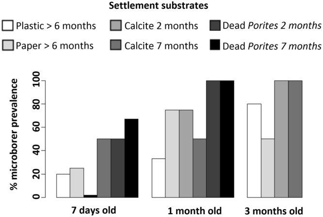
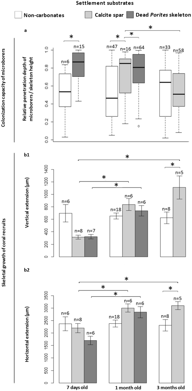
Ostreobium sp. (Bryopsidales, Ulvophyceae) is a major microboring alga involved in tropical reef dissolution, with a proposed symbiotic lifestyle in living corals. However, its diversity and colonization dynamics in host's early life stages remained unknown. Here, we mapped microborer distribution and abundance in skeletons of the branching coral Pocillopora damicornis from the onset of calcification in primary polyps (7 days) to budding juvenile colonies (1 and 3 months) growing on carbonate and non-carbonate substrates pre-colonized by natural biofilms, and compared them to adult colonies (in aquarium settings). Primary polyps were surprisingly already colonized by microboring filaments and their level of invasion depended on the nature of settlement substrate and the extent of its pre-colonization by microborers. Growth of early coral recruits was unaffected even when microborers were in close vicinity to the polyp tissue. In addition to morphotype observations, chloroplast-encoded rbcL gene sequence analyses revealed nine new Ostreobium clades (OTU99%) in Pocillopora coral. Recruits and adults shared one dominant rbcL clade, undetected in larvae, but also present in aquarium seawater, carbonate and non-carbonate settlement substrates, and in corals from reef settings. Our results show a substratum-dependent colonization by Ostreobium clades, and indicate horizontal transmission of Ostreobium-coral associations.


Conflict of interest statement

The authors declare that they have no competing interests.

Figures

Figure 1
Figure 1
Filamentous microborers observed in coral skeletons. Light (a, c, e) and scanning electron micrographs of the associated microborings or galleries (b, d, f). (a) Phaeophila sp. in dead Porites substrate stained with toluidine blue (black arrow). (b) The corresponding resin- replicated galleries (white arrow). (c) Ostreobium sp. in 1 month old P. damicornis recruit stained with Grocott’s Methenamine Silver (black arrow). (d) The corresponding galleries in dead Porites substrate (white arrow). (e) Fungal hyphae in adult P. damicornis colony stained with toluidine blue (black arrow). (f) The corresponding galleries in dead Porites substrate (white arrow): fungi and Ostreobium galleries* are in close proximity.
Figure 2
Figure 2
Substratum-dependent microborer colonization of coral recruits. Euendolithic filaments were detected by photonic and/or scanning electron microscopy in skeletons of P. damicornis (2 to 9 replicates per life stage and substrate type). One month old recruits developed on clean glass had 25% microborer prevalence. Data for 3 months old recruits on dead Porites are missing because full colonization was already reached on these substrates in 1 month old recruits (100% prevalence) and -given the limited availability of larval material- focus was given to the colonization process.
Figure 3
Figure 3
Colonization dynamics of skeletons of coral recruits and adult colonies by microborers. (a) One month old P. damicornis recruit. (b1) Thin section and (b2) corresponding distribution/abundance mapping of microborers in one month old recruit settled on non-carbonate (plastic) or (c1,c2) carbonate substrate (dead Porites skeleton). (d, e1, f1) Adult P. damicornis colony branch. (e2) Thin section and (e3) corresponding distribution/abundance mapping of microborers in an adult colony; (f2, f3) also illustrated for another adult sample. The higher is the abundance, the warmer is the color code on maps. Dotted line represents the boundary between tissue-covered skeleton (apex) and dead skeletal base. Orientation of thin sections is parallel to coral vertical growth axis (dark blue arrow).
Figure 4
Figure 4
Microborer penetration into rapidly extending skeletons of early coral recruits. (a) The colonization capacity is reported as relative depth of penetration of microborers to skeletal height (relative DP) and varies among larval settlement substrates; n = number of measurements of depths of penetration. (b1) Corresponding vertical and (b2) horizontal extension rates of n replicate coral recruits, with standard error. *Significant differences (P < 0.05). Data for 3 months old recruits on dead Porites are missing because monitoring of larval development on these substrates was stopped at 1 month when prevalence of microborers in coral skeleton reached 100% (see Fig. 2).

References

    1. Rosenberg E, Koren O, Reshef L, Efrony R, Zilber-Rosenberg I. The role of microorganisms in coral health, disease and evolution. Nat. Rev. Microbiol. 2007;5:355–362. doi: 10.1038/nrmicro1635. - DOI - PubMed
    1. Tribollet, A. The boring microflora in modern coral reef ecosystems: a review of its roles. In Curr. Dev. Bioerosion (eds Wisshak, M. & Tapanila, L.) 67–94 (Springer Berlin Heidelberg, 2008).
    1. Marcelino, V. R. & Verbruggen, H. Multi-marker metabarcoding of coral skeletons reveals a rich microbiome and diverse evolutionary origins of endolithic algae. Sci. Rep. 6, 31508 (2016). - PMC - PubMed
    1. Halldal P. Photosynthetic capacities and photosynhtetic action spectra of endozoic algae of the massive coral. Favia. Biol. Bull. 1968;134:411–424. doi: 10.2307/1539860. - DOI
    1. Shibata K, Haxo FT. Light transmission and spectral distribution through epi- and endozoic algal layers in the brain coral. Favia. Biol. Bull. 1969;136:461–468. doi: 10.2307/1539688. - DOI

Publication types

Substances

LinkOut - more resources