. 2018 Feb;35(1-2):37-51.
doi: 10.1007/s10585-018-9876-z. Epub 2018 Feb 2.

Pharmacologic ascorbate (P-AscH-) suppresses hypoxia-inducible Factor-1α (HIF-1α) in pancreatic adenocarcinoma

Affiliations

Pharmacologic ascorbate (P-AscH-) suppresses hypoxia-inducible Factor-1α (HIF-1α) in pancreatic adenocarcinoma

Justin G Wilkes et al. Clin Exp Metastasis. 2018 Feb.

Abstract

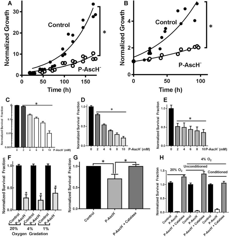
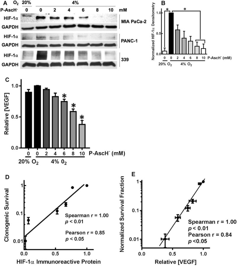
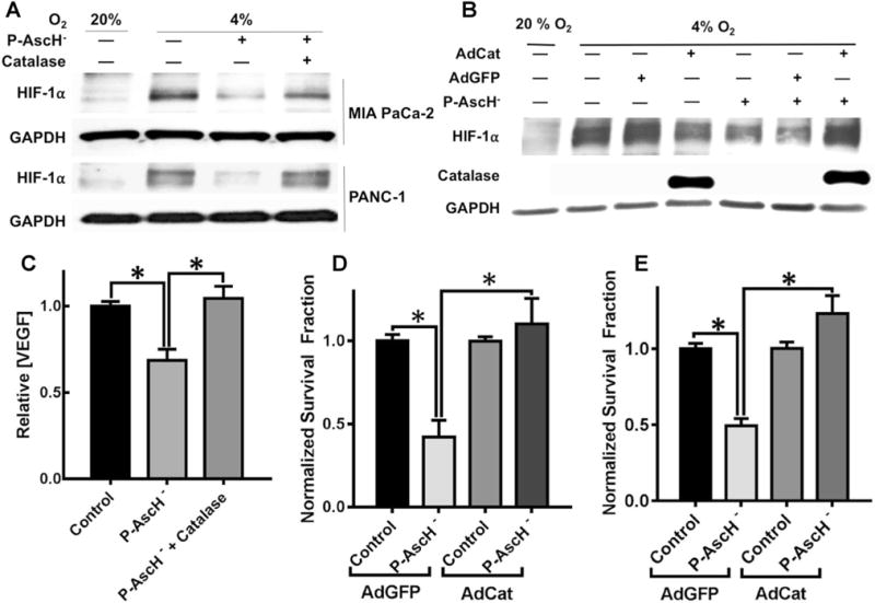
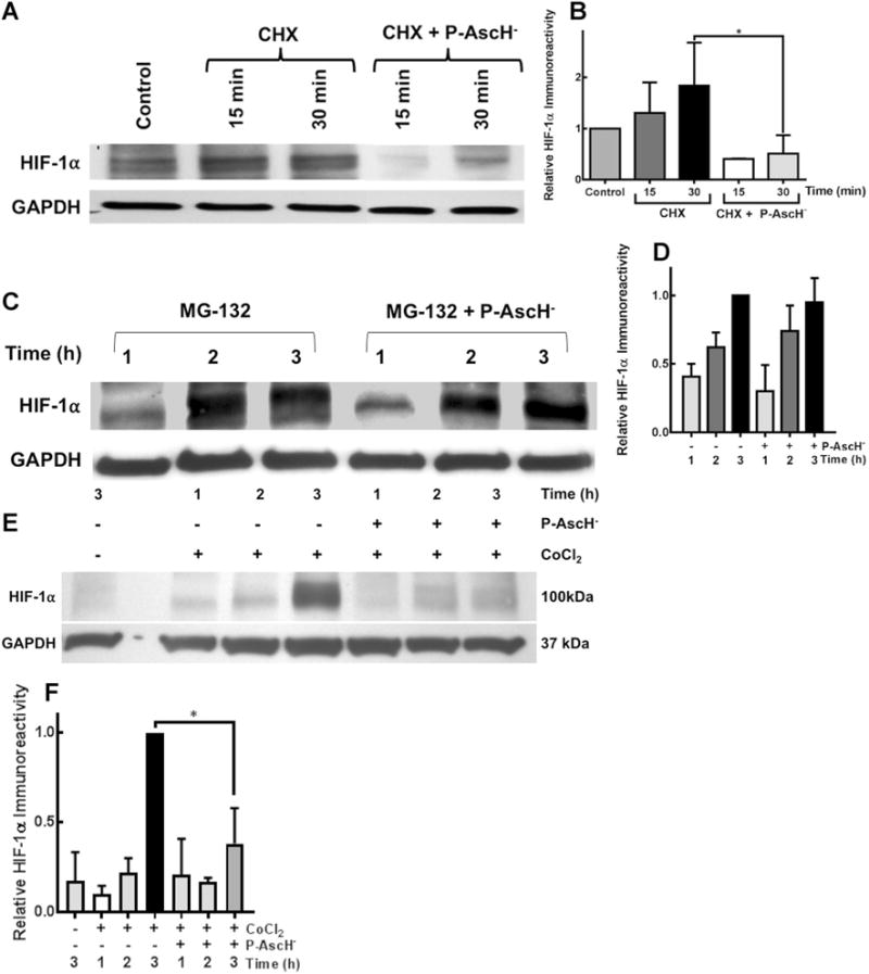
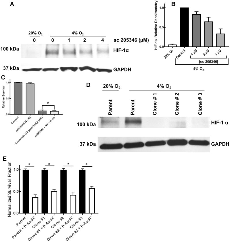
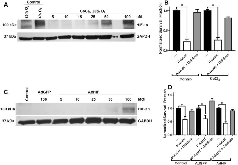
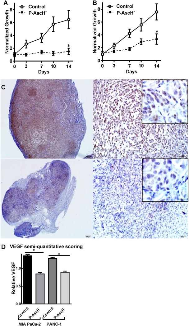
HIF-1α is a transcriptional regulator that functions in the adaptation of cells to hypoxic conditions; it strongly impacts the prognosis of patients with cancer. High-dose, intravenous, pharmacological ascorbate (P-AscH-), induces cytotoxicity and oxidative stress selectively in cancer cells by acting as a pro-drug for the delivery of hydrogen peroxide (H2O2); early clinical data suggest improved survival and inhibition of metastasis in patients being actively treated with P-AscH-. Previous studies have demonstrated that activation of HIF-1α is necessary for P-AscH- sensitivity. We hypothesized that pancreatic cancer (PDAC) progression and metastasis could be be targeted by P-AscH- via H2O2-mediated inhibition of HIF-1α stabilization. Our study demonstrates an oxygen- and prolyl hydroxylase-independent regulation of HIF-1α by P-AscH-. Additionally, P-AscH- decreased VEGF secretion in a dose-dependent manner that was reversible with catalase, consistent with an H2O2-mediated mechanism. Pharmacological and genetic manipulations of HIF-1α did not alter P-AscH--induced cytotoxicity. In vivo, P-AscH- inhibited tumor growth and VEGF expression. We conclude that P-AscH- suppresses the levels of HIF-1α protein in hypoxic conditions through a post-translational mechanism. These findings suggest potential new therapies specifically designed to inhibit the mechanisms that drive metastases as a part of PDAC treatment.

Keywords: Ascorbate; Hypoxia inducible factor; Metastasis; Pancreatic adenocarcinoma; Vitamin C.


Conflict of interest statement

Conflict of interest The Authors declare that they have no conflicts of interest with the contents of this article.

Figures

Fig. 1
Fig. 1
P-AscH-induced cytotoxicity in hypoxia. a Treatment of MIA PaCa-2 cells with P-AscH (7 mM; 7 pmol cell−1) significantly increased doubling time (n = 6, *p < 0.05 vs. control). b P-AscH (10 mM; 10 pmol cell−1) significantly increased doubling time in PANC-1 cancer cell lines (n = 6, p < 0.05). c Clonogenic survival was decreased in a dose-dependent fashion in MIA-PaCa-2 cells treated and subsequently grown in 4% O2 (n = 3, *p < 0.01 vs. control). d Clonogenic survival was decreased in a dose-dependent fashion in PANC-1 cells treated and subsequently grown in 4% O2 (n = 3, *p < 0.01 vs. control). e Clonogenic survival was decreased in a dose-dependent fashion in 339 cells treated and subsequently grown in 4% O2 (n = 4, *p < 0.01 vs. control). f P-AscH-induced decreases in clonogenic survival were similar in hypoxic environments (7 pmol/cell, 7 mM P-AscH, means ± SEM, n = 3, *p < 0.05 vs. control). g Catalase reverses P-AscH-induced cytotoxicity in 1% O2 (7 pmol/cell, 7 mM P-AscH, means ± SEM, n = 3, *p < 0.05 vs. control). h Clonogenic survival of P-AscH treated cells pre-conditioned to hypoxia by 16-h incubation in 4% O2 compared to unconditioned cells and cells treated in 20% O2. Cells conditioned to hypoxia were significantly sensitive to P-AscH (7 pmol/cell, 7 mM P-AscH, n = 3, p < 0.01), however, pre-conditioning to hypoxia yielded no significant resistance to P-AscH relative to unconditioned controls, or cells treated in 20% O2
Fig. 2
Fig. 2
P-AscH suppresses HIF-1α immunoreactive protein and VEGF secretion. a MIA PaCa-2, PANC-1, and 339 cell lines showed a dose-dependent decrease in HIF-1α expression after 1-h exposure to P-AscH. Both cell lines had induction of HIF-1α immunoreactive protein in hypoxia. b In the MIA PaCa-2 cell line, densitometric analysis of HIF-1α demonstrates a decrease in HIF-1α expression with increasing doses of P-AscH (n = 3, p < 0.05). c Secreted VEGF in media after 1-h P-AscH and incubation in 4% O2 also demonstrated a dose-dependent decrease (means ± SEM, n = 6, *p < 0.05). d Clonogenic survival correlated with HIF-1α immunoreactive protein in MIA PaCa-2 cancer cells treated with P-AscH (2–10 mM). e Clonogenic survival correlated with VEGF secretion in MIA PaCa-2 cancer cells with P-AscH (2–10 mM)
Fig. 3
Fig. 3
Catalase reverses the hypoxic P-AscH-induced suppression of clonogenic survival, HIF-1α and VEGF. a Western Blot demonstrates the reversal of P-AscH-induced (2 mM, 5.4 pmol cell−1) decrease in HIF-1α in 4% O2 with extracellular catalase in both MIA PaCa-2 and PANC-1 cell lines. Again, hypoxia (4% O2) induced HIF-1α in both cell lines. P-AscH decreased HIF-1α immunoreactive protein which was then reversed with addition of exogenous catalase. b Over-expression of intracellular catalase reverses the P-AscH-induced (2 mM, 5.4 pmol cell−1) HIF-1α suppression. HIF-1α was induced with hypoxia while cells were infected with an adenovirus containing the cDNA for catalase or containing GFP (control vector). Catalase immunoreactivity demonstrated robust increase in catalase protein with the AdCat vector. Catalase overexpression had no effect of HIF-1α in hypoxia; however, catalase overexpression reversed the P-AscH-induced HIF-1α suppression. c P-AscH-induced decreases in VEGF secretion were reversed with extracellular scavenging of H2O2 by catalase (MIA PaCa-2, n = 6, *p < 0.05, Mean ± SEM). d P-AscH-induced (5 mM, 7 pmol cell−1) clonogenic cell death which was reversed with overexpression of intracellular catalase in MIA PaCa-2 cells (means ± SEM, n = 3, *p < 0.05). e P-AscH-induced (5 mM, 7 pmol cell−1) clonogenic cell death was reversed with over-expression of intracellular catalase in PANC-1cells (means ± SEM, n = 3, *p < 0.05)
Fig. 4
Fig. 4
P-AscH-induced HIF-1α suppression is due to a prolyl hydroxylase-independent increase in degradation. a Ribosomal inhibition in hypoxia with cycloheximide (CHX) and subsequent treatment with P-AscH (5 mM, 7 pmol cell− 1) results in decreased HIF-1α as demonstrated by Western blot. b Densitometric analysis demonstrates decreased HIF-1α with CHX and P-AscH (*p < 0.1, n = 3, One-Way ANOVA, Dunn’s multiple comparisons test). c Proteasomal inhibition utilizing MG-132 reverses P-AscH induced (5 mM, 7 pmol cell−1) suppression of HIF-1α protein as demonstrated by Western blot. d Densitometric analysis, suggesting P-AscH increases degradation of HIF-1α as opposed to inhibiting synthesis. e Representative Western blot demonstrating inhibition of prolyl hydroxylase utilizing cobalt chloride (CoCl2) fails to reverse P-AscH-induced (7 mM, 7 pmol cell−1) HIF-1α protein suppression. f Densitometric analysis demonstrated that CoCl2 does not reverse P-AscH HIF-1α suppression (*p < 0.05, n = 3)
Fig. 5
Fig. 5
Selective HIF-1α suppression does not reverse P-AscH-induced cytotoxicity. a Western blotting demonstrates induction of HIF-1α protein in hypoxia. Pharmacologic inhibition with sc-205346 suppressed HIF-1α as demonstrated by Western blotting. b Densitometry from Western blots demonstrating decreasing HIF-1α with pharmacologic inhibition using the compound sc-205356. c Pharmacological inhibition of HIF-1α does not alter P-AscH (6 mM, 12 pmol cell−1) decreases in clonogenic survival (means ± SEM, n = 3 * p < 0.05 vs. control). d Three clones of MIA PaCa-2 cancer cells subjected to CRISPR/Cas9 HIF-1α genome editing demonstrated decreased expression of HIF-1α in hypoxia (4% O2, 8 h). e Decreased HIF-1α expression did not alter P-AscH (10 mM) induced decreases in clonogenic survival (means ± SEM, *p < 0.0001 comparing each respective untreated cell line vs. P-AscH treated cell lines. The untreated cell lines were normalized to 1.0, n = 3)
Fig. 6
Fig. 6
HIF-1α overexpression confers no resistance to P-AscH. a Western blot of protein collected from MIA PaCa-2 cells incubated in normoxia with increasing concentration of CoCl2 (5–100 μM). Significant increases in HIF-1α protein are demonstrated at 50–100 μM CoCl2. b Clonogenic survival of MIA PaCa-2 cells pre-incubated for 8 h with CoCl2 (50 μM) prior to treatment with P-AscH for 1 h demonstrates significant cytotoxicity (n = 3, p < 0.01) relative to non-P-AscH controls. There were no significant differences in survival between P-AscH-treated cells incubated with CoCl2 compared to untreated cells. c Western blot of protein collected from MIA PaCa-2 cells transfected with increasing MOI of AdHIF adenovirus. Significant overexpression of HIF-1α was demonstrated at 100 MOI. d Clonogenic survival of MIA PaCa-2 cells transfected with AdHIF adenovirus (100 MOI) treated with P-AscH for 1 h in hypoxia. P-AscH induced a significant cytotoxicity to AdHIF transfected cells (n = 3, p < 0.01) that was not significantly different from AdGFP and non-transfected controls
Fig. 7
Fig. 7
P-AscH inhibits in vivo tumor growth and VEGF expression. A MIA PaCa-2 human tumor xenografts in athymic nude mice have significantly decreased tumor growth in mice treated with daily I.P. P-AscH (4 g kg−1, b.i.d.) compared to mice receiving saline (control). Comparison of normalized mean tumor size at 14 days demonstrates significantly decreased growth of control mice and P-AscH-treated mice (1.5-fold relative growth vs. 6.5-fold relative growth at 2 weeks, means ± SEM, n = 8, *p < 0.05). b Comparison of normalized mean tumor growth at 14 days demonstrated significantly decreased growth in P-AscH treated mice compared to control mice in PANC-1 xenografts (3.7-fold relative growth vs. 9.1-fold relative growth at 2 weeks, means ± SEM, n = 8, *p < 0.05.). C. Representative VEGF immunohistochemistry (brown cytoplasmic staining) of MIA PaCa-2 tumor xenograft treated with saline (top panels) and P-AscH (bottom panels). d Immunohistochemistry for VEGF. Sections were incubated with polyclonal rabbit anti-VEGF. VEGF stained slides were blindly evaluated by a board-certified veterinary pathologist (KGC) and graded using a semi-quantitative scoring system on a 0–3 scale. Twenty separate fields of view per slide were evaluated and analyzed. VEGF Semi-quantitative immunohistochemistry of all xenografts demonstrate significant decreased VEGF expression (n = 8, means ± SEM, *p < 0.01) in tumors of mice treated with P-AscH

References

    1. Matsuo Y, Ding Q, Desaki R, et al. Hypoxia inducible factor-1 alpha plays a pivotal role in hepatic metastasis of pancreatic cancer: an immunohistochemical study. J Hepatobiliary Pancreat Sci. 2014;21(2):105–112. - PubMed
    1. Sun HC, Qiu ZJ, Liu J, et al. Expression of hypoxia-inducible factor-1 alpha and associated proteins in pancreatic ductal adenocarcinoma and their impact on prognosis. Int J Oncol. 2007;30(6):1359–1367. - PubMed
    1. Hoffmann AC, Mori R, Vallbohmer D, et al. High expression of HIF1a is a predictor of clinical outcome in patients with pancreatic ductal adenocarcinomas and correlated to PDGFA, VEGF, and bFGF. Neoplasia. 2008;10(7):674–679. - PMC - PubMed
    1. Shibaji T, Nagao M, Ikeda N, et al. Prognostic significance of HIF-1 alpha overexpression in human pancreatic cancer. Anticancer Res. 2003;23(6C):4721–4727. - PubMed
    1. Buchler P, Reber HA, Buchler M, et al. Hypoxia-inducible factor 1 regulates vascular endothelial growth factor expression in human pancreatic cancer. Pancreas. 2003;26(1):56–64. - PubMed

Publication types

MeSH terms

Substances

LinkOut - more resources