. 2018 Feb;136(1):1-26.
doi: 10.1007/s10633-017-9621-y. Epub 2018 Feb 3.

ISCEV guide to visual electrodiagnostic procedures

Affiliations

ISCEV guide to visual electrodiagnostic procedures

Anthony G Robson et al. Doc Ophthalmol. 2018 Feb.

Abstract

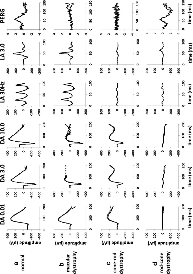
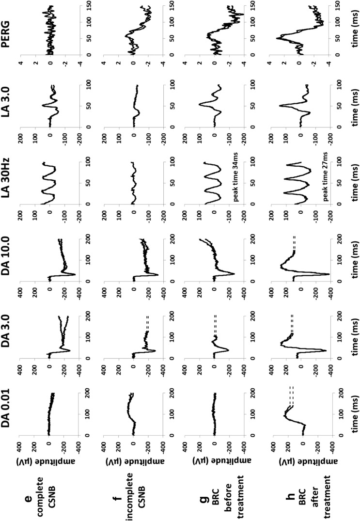
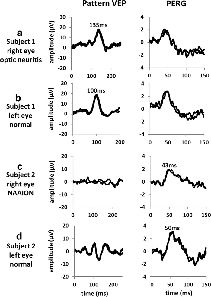
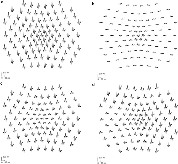
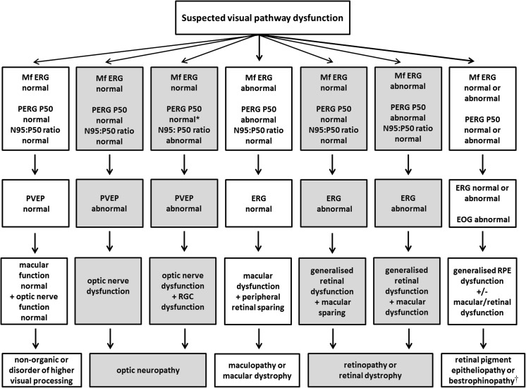
Clinical electrophysiological testing of the visual system incorporates a range of noninvasive tests and provides an objective indication of function relating to different locations and cell types within the visual system. This document developed by the International Society for Clinical Electrophysiology of Vision provides an introduction to standard visual electrodiagnostic procedures in widespread use including the full-field electroretinogram (ERG), the pattern electroretinogram (pattern ERG or PERG), the multifocal electroretinogram (multifocal ERG or mfERG), the electrooculogram (EOG) and the cortical-derived visual evoked potential (VEP). The guideline outlines the basic principles of testing. Common clinical presentations and symptoms are described with illustrative examples and suggested investigation strategies.

Keywords: Clinical electrophysiology; Electrooculogram (EOG); Electroretinogram (ERG); ISCEV standards; Maculopathy; Multifocal ERG (mfERG); Optic neuropathy; Pattern ERG; Retinopathy; Visual evoked potential (VEP).


Conflict of interest statement

Conflict of interest

All authors certify that they have no affiliations with or involvement in any organization or entity with any financial interest (such as honoraria; educational grants; participation in speakers’ bureaus; membership, employment consultancies, stock ownership, or other equity interest; and expert testimony or patent-licensing arrangements) or non-financial interest (such as personal or professional relationships, affiliations, knowledge or beliefs) in the subject matter or materials discussed in this manuscript.

Informed consent

For this type of study formal consent is not required.

Statement of human rights

This article does not contain any research studies with human participants performed by any of the authors.

Statement on the welfare of animals

This article does not contain any research studies with animals performed by any of the authors.

Figures

Fig. 1
Fig. 1
Representative full-field and pattern ERGs in a normal subject (a), in a case of macular dystrophy (b), cone-rod dystrophy (c), rod-cone dystrophy with relative sparing of macular function (d), complete CSNB (e), incomplete CSNB (f) and birdshot retinochoroidopathy (BRC) before treatment (g) and after treatment illustrating full recovery of the ERG and PERG (h). Recordings showed a high degree of inter-ocular symmetry except in BRC (data from other eye are not shown). Note there is a 20-ms pre-stimulus delay in all single flash ERG recordings. Two responses for each stimulus condition are superimposed to illustrate reproducibility. Broken lines replace blink artefacts occurring after the ERGs, for clarity
Fig. 1
Fig. 1
Representative full-field and pattern ERGs in a normal subject (a), in a case of macular dystrophy (b), cone-rod dystrophy (c), rod-cone dystrophy with relative sparing of macular function (d), complete CSNB (e), incomplete CSNB (f) and birdshot retinochoroidopathy (BRC) before treatment (g) and after treatment illustrating full recovery of the ERG and PERG (h). Recordings showed a high degree of inter-ocular symmetry except in BRC (data from other eye are not shown). Note there is a 20-ms pre-stimulus delay in all single flash ERG recordings. Two responses for each stimulus condition are superimposed to illustrate reproducibility. Broken lines replace blink artefacts occurring after the ERGs, for clarity
Fig. 2
Fig. 2
Representative pattern-reversal VEPs and PERGs in the affected (a, c) and fellow (b, d) eyes in a patient with non-acute optic neuritis (Subject 1; a, b) and in an elderly patient with a severe non-arteritic anterior ischemic optic neuropathy (Subject 2; c, d). The P100 component of the pattern VEP in optic neuritis shows a 35-ms delay compared with the normal fellow eye, without significant amplitude reduction, consistent with optic nerve conduction delay; pattern ERGs are normal in this case and reveal no evidence of macular or retinal ganglion cell dysfunction. The pattern VEP P100 component in c is undetectable, and PERG shows a reduced N95:P50 ratio and shortening of P50 peak time (inter-ocular difference 7 ms) compared with the fellow eye, indicating severe optic nerve dysfunction with retinal ganglion cell involvement. Two responses for each stimulus condition are superimposed to illustrate reproducibility
Fig. 3
Fig. 3
Multifocal ERGs recorded to a 103-element stimulus array in a representative normal subject (a), in a case of retinitis pigmentosa showing relative sparing of central macular function (b), in a case of macular dystrophy showing reduction over a central area (c) and in a patient with an eccentric nasal area of retinal dysfunction consistent with an enlarged blind spot extending inferiorly in the right eye (d). MfERGs in cases ac showed a high degree of inter-ocular symmetry; abnormalities were unilateral in d. Traces are shown in retinal view
Fig. 4
Fig. 4
Suggested test strategy for cases of suspected visual pathway dysfunction, illustrating how complementary tests can localize dysfunction within the visual system. Asterisk (*): in cases of retinal ganglion cell dysfunction, the PERG N95:P50 ratio is subnormal, but in severe disease P50 may additionally show reduction with shortening of peak time. Dagger (†): bestrophinopathies; Best disease is associated with a normal ERG and abnormal EOG; autosomal recessive bestrophinopathy causes severe EOG reduction and later onset progressive retinopathy with relatively mild ERG abnormality; in ADVIRC, the EOG is abnormal and the ERG abnormal. See Table 1 for details. After; [6, 7]

References

    1. McCulloch DL, Marmor MF, Brigell MG, Hamilton R, Holder GE, Tzekov R, Bach M. ISCEV standard for full-field clinical electroretinography (2015 update) Doc Ophthalmol. 2015;130:1–12. doi: 10.1007/s10633-014-9473-7. - DOI - PubMed
    1. Bach M, Brigell MG, Hawlina M, Holder GE, Johnson MA, McCulloch DL, Meigen T, Viswanathan S. ISCEV standard for clinical pattern electroretinography (PERG): 2012 update. Doc Ophthalmol. 2013;126:1–7. doi: 10.1007/s10633-012-9353-y. - DOI - PubMed
    1. Constable PA, Bach M, Frishman LJ, Jeffrey BG, Robson AG. International society for clinical electrophysiology of vision. ISCEV standard for clinical electro-oculography (2017 update) Doc Ophthalmol. 2017;134:1–9. doi: 10.1007/s10633-017-9573-2. - DOI - PMC - PubMed
    1. Hood DC, Bach M, Brigell M, Keating D, Kondo M, Lyons JS, Marmor MF, McCulloch DL, Palmowski-Wolfe AM. ISCEV standard for clinical multifocal electroretinography (mfERG) (2011 edition) Doc Ophthalmol. 2012;124:1–13. doi: 10.1007/s10633-011-9296-8. - DOI - PMC - PubMed
    1. Odom JV, Bach M, Brigell M, Holder GE, McCulloch DL, Mizota A, Tormene AP. ISCEV standard for clinical visual evoked potentials—(2016 update) Doc Ophthalmol. 2016;133(1):1–9. doi: 10.1007/s10633-016-9553-y. - DOI - PubMed

Publication types

MeSH terms

LinkOut - more resources