. 2018 Aug;24(15-16):1218-1227.
doi: 10.1089/ten.TEA.2017.0438. Epub 2018 Apr 23.

ROCK Inhibition Promotes the Development of Chondrogenic Tissue by Improved Mass Transport

Affiliations

ROCK Inhibition Promotes the Development of Chondrogenic Tissue by Improved Mass Transport

Kuo-Chen Wang et al. Tissue Eng Part A. 2018 Aug.

Abstract

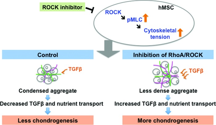
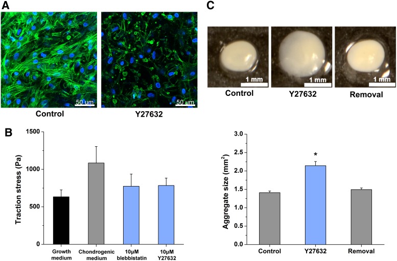
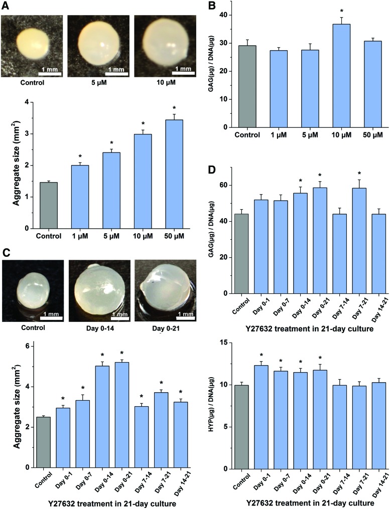
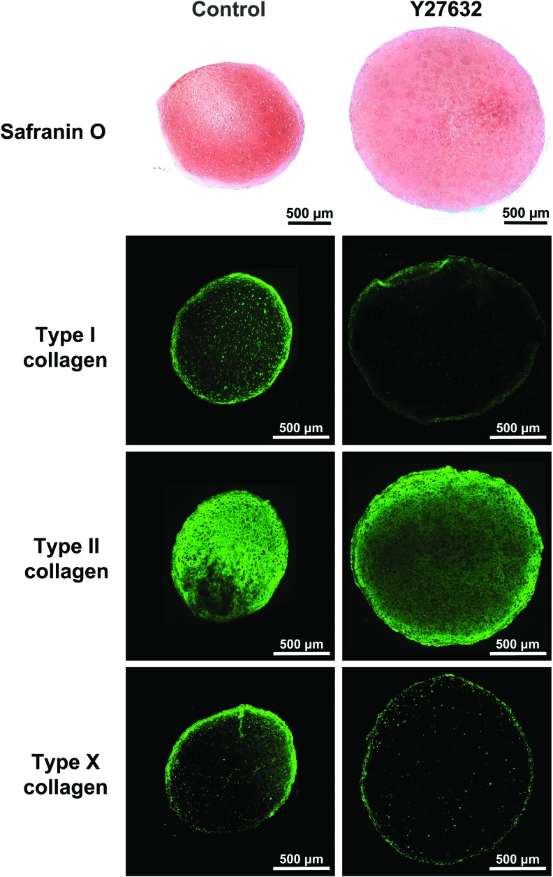
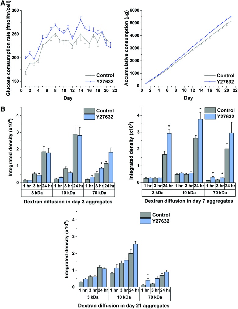
Human mesenchymal stem cell (hMSC)-based chondrogenesis is a key process used to develop tissue engineered cartilage constructs from stem cells, but the resulting constructs have inferior biochemical and biomechanical properties compared to native articular cartilage. Transforming growth factor β containing medium is commonly applied to cell layers of hMSCs, which aggregate upon centrifugation to form 3-D constructs. The aggregation process leads to a high cell density condition, which can cause nutrient limitations during long-term culture and, subsequently, inferior quality of tissue engineered constructs. Our objective is to modulate the aggregation process by targeting RhoA/ROCK signaling pathway, the chief modulator of actomyosin contractility, to enhance the end quality of the engineered constructs. Through ROCK inhibition, repression of cytoskeletal tension in chondrogenic hMSCs was achieved along with less dense aggregates with enhanced transport properties. ROCK inhibition also led to significantly increased cartilaginous extracellular matrix accumulation. These findings can be used to create an improved microenvironment for hMSC-derived tissue engineered cartilage culture. We expect that these findings will ultimately lead to improved cartilaginous tissue development from hMSCs.

Keywords: chondrogenesis; human mesenchymal stem cells; mass transport; signaling; tissue engineering.


Conflict of interest statement

No competing financial interests exist.

Figures

<b>FIG. 1.</b>
FIG. 1.
Hypothesized effect of RhoA/ROCK signaling inhibition on the development of hMSC-based chondrogenic tissue. The chondrogenic aggregate under RhoA/ROCK signaling inhibition is hypothesized to be loosely formed through downregulation of cytoskeletal tension. The transport property within the tissue is therefore enhanced and that results in improvement in cartilaginous tissue development. (Lines in gray: hMSCs, in pink: glycosaminoglycan [GAG], in green: collagen.) hMSCs, human mesenchymal stem cells; TGFβ, transforming growth factor β. Color images available online at www.liebertpub.com/tea
<b>FIG. 2.</b>
FIG. 2.
Inhibition of RhoA/ROCK signaling attenuates hMSC cytoskeleton tension and compactness of chondrogenic aggregation. (A) Evaluation of hMSC stress fiber formation under control and 10 μM Y27632 treatment during chondrogenic induction. Phalloidin (green) and DAPI (blue) were used for stress fiber and nuclear staining, respectively, and imaged at 50 μm depth from the surface of control and 10 μM Y27632 treated intact aggregates at Day 7. Scale bar 50 μm. (n = 8) (B) The change of contraction force generated by hMSCs in growth medium (control), chondrogenic differentiation medium, and chondrogenic medium with RhoA/ROCK signaling inhibitors, 10 μM blebbistatin or Y27632. (n ≥ 37) Student's t-test results for comparison to growth maintenance (control): chondrogenic differentiation medium p = 0.06; 10 μM blebbistatin p = 0.26; 10 μM Y27632 p = 0.22. (C) Assessment of the effect of 10 μM Y27632 treatment on hMSC chondrogenic aggregation. Images of aggregates at 48 h (top panel): untreated (left, control) and 10 μM Y27632 treated (middle, Y27632) aggregates. The aggregate on the right (removal) was exposed to 10 μM Y27632 treatment for the first 24 h only. Scale bar 1 mm. Bottom graph shows image analysis data. (n = 10) Data represent the mean ± SEM. ANOVA F-test; *p < 0.05. See also Supplementary Figure S1. ANOVA, analysis of variance; DAPI, 4′,6-diamidino-2-phenylindole dihydrochloride. Color images available online at www.liebertpub.com/tea
<b>FIG. 3.</b>
FIG. 3.
Effect of dosage and exposure regime of Y27632 treatment on hMSC chondrogenic differentiation. (A) Morphological properties of hMSC aggregates after Y27632 treatment. Aggregates of control and Y27632 treatment at 1, 5, 10, or 50 μM were harvested after 21-day culture. Estimated size of aggregates was obtained from the projected areas of the images. Scale bar 1 mm. (n = 8) (B) Evaluation of Y27632 dosage effect on GAG accumulation after 21-day culture. The amount of GAG in digested aggregates was measured and normalized to DNA content for comparison. (n ≥ 8) (C) Morphological properties of hMSC aggregates after 10 μM Y27632 treatment in different periods of 21-day culture. The aggregates were imaged after harvest for size measurements. Scale bar 1 mm. (n ≥ 11) (D) The effect of different exposure regimes of 10 μM Y27632 on GAG and HYP depositions. The quantities of GAG and HYP in digested hMSC aggregates were normalized to DNA content to yield the result of GAG per DNA and HYP per DNA for comparison. (n ≥ 11) Data represent the mean ± SEM. ANOVA F-test; *p < 0.05. See also Supplementary Figures S2–S5. Color images available online at www.liebertpub.com/tea
<b>FIG. 4.</b>
FIG. 4.
Histology and immunohistochemistry results. Safranin-O and type I, II, and X collagen stained hMSC aggregates cultured with 10 μM Y27632 for 21 days were compared with control. Scale bar 500 μm. (n ≥ 4) Color images available online at www.liebertpub.com/tea
<b>FIG. 5.</b>
FIG. 5.
Assessment of transport properties in hMSC chondrogenic aggregates under Y27632 treatment during 21-day culture. (A) Glucose consumption in hMSC aggregates after 10 μM Y27632 treatment. Daily glucose consumption rate and accumulative glucose consumption in 21-day culture were analyzed. (n = 6) (B) Diffusional status of hMSC aggregates under ROCK inhibition at different chondrogenic stages. The intensities of tissue sections in control and 10 μM Y27632 treated aggregates exposed to 3, 10, and 70 kDa fluorescent dextran tracers for 1, 3, and 24 h at day 3, 7, and 21 culture times were analyzed. (n ≥ 4) Data represent the mean ± SEM. ANOVA F-test; *p < 0.05. See also Supplementary Table S1. Color images available online at www.liebertpub.com/tea

Similar articles

Cited by

References

    1. Pittenger M.F., Mackay A.M., Beck S.C., et al. . Multilineage potential of adult human mesenchymal stem cells. Science 284, 143, 1999 - PubMed
    1. Fell H.B. The histogenesis of cartilage and bone in the long bones of the embryonic fowl. J Morphol 40, 417, 1925
    1. Johnstone B., Hering T.M., Caplan A.I., Goldberg V.M., and Yoo J.U. In vitro chondrogenesis of bone marrow-derived mesenchymal progenitor cells. Exp Cell Res 238, 265, 1998 - PubMed
    1. Yoo J.U., Barthel T.S., Nishimura K., et al. . The chondrogenic potential of human bone-marrow-derived mesenchymal progenitor cells. J Bone Joint Surg Am 80, 1745, 1998 - PubMed
    1. Hillel A.T., Taube J.M., Cornish T.C., et al. . Characterization of human mesenchymal stem cell-engineered cartilage: analysis of its ultrastructure, cell density and chondrocyte phenotype compared to native adult and fetal cartilage. Cells Tissues Organs 191, 12, 2010 - PubMed

Publication types

Substances

LinkOut - more resources