. 2018 Apr 19;25(4):392-402.e14.
doi: 10.1016/j.chembiol.2018.01.006. Epub 2018 Feb 1.

CD1b Tetramers Identify T Cells that Recognize Natural and Synthetic Diacylated Sulfoglycolipids from Mycobacterium tuberculosis

Affiliations

CD1b Tetramers Identify T Cells that Recognize Natural and Synthetic Diacylated Sulfoglycolipids from Mycobacterium tuberculosis

Charlotte A James et al. Cell Chem Biol. .

Abstract

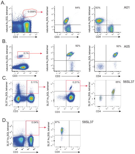
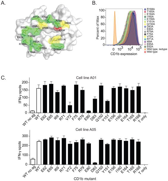
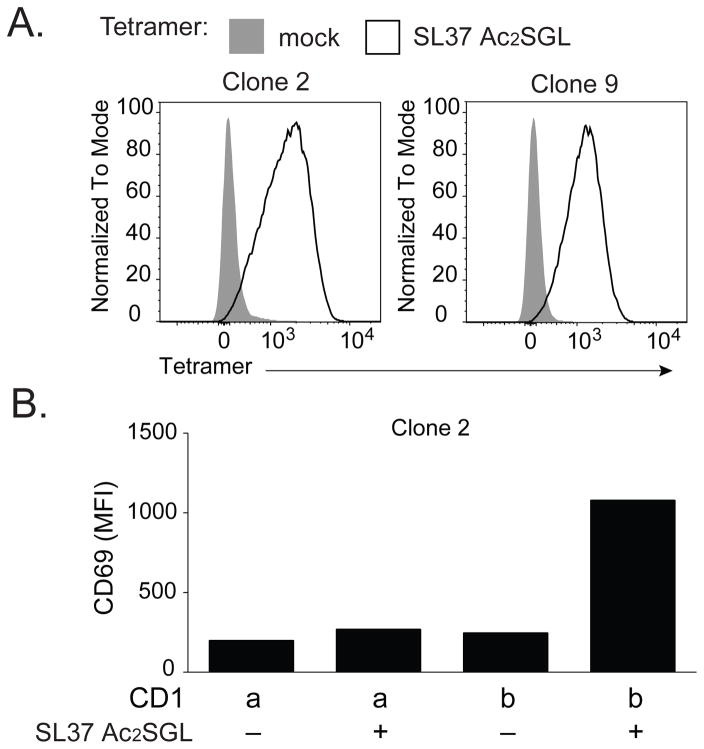
Mycobacterial cell wall lipids bind the conserved CD1 family of antigen-presenting molecules and activate T cells via their T cell receptors (TCRs). Sulfoglycolipids (SGLs) are uniquely synthesized by Mycobacterium tuberculosis, but tools to study SGL-specific T cells in humans are lacking. We designed a novel hybrid synthesis of a naturally occurring SGL, generated CD1b tetramers loaded with natural or synthetic SGL analogs, and studied the molecular requirements for TCR binding and T cell activation. Two T cell lines derived using natural SGLs are activated by synthetic analogs independently of lipid chain length and hydroxylation, but differentially by saturation status. By contrast, two T cell lines derived using an unsaturated SGL synthetic analog were not activated by the natural antigen. Our data provide a bioequivalence hierarchy of synthetic SGL analogs and SGL-loaded CD1b tetramers. These reagents can now be applied to large-scale translational studies investigating the diagnostic potential of SGL-specific T cell responses or SGL-based vaccines.

Keywords: CD1; T cell receptor; T cells; antigen-presentation; human; lipid antigen; mycobacteria; tuberculosis.


Figures

Fig. 2
Fig. 2
Structures of Hydroxyphthioceranic acid (1) and Phthioceranic acid (2)
Figure 1
Figure 1. Diacylated sulfoglycolipid (Ac2SGL) antigens
(A) Structures of natural Ac2SGL purified from M.tb and three synthetic forms that have been previously described: SL37 Ac2SGL, SL27 Ac2SGL, SL29 Ac2SGL. Synthesis of a fourth analog (AM Ac2SGL) is reported here. These compounds are used to probe the specificity of T-cell responses to Ac2SGL antigens. (B) Hybrid synthesis method for hydroxyphthioceranic acid consisting of an iterative conjugated addition and lithiation/borylation. TBDPS = tert-butyldiphenylsilyl, MOM = methoxymethyl, Pin = pinacolato. (C) Regioselective addition of hydroxyphthioceranoic acid to the trehalose core followed by 2′-O-sulfation and deprotection to yield AM Ac2SGL. Validation of the compounds by mass and NMR spectrometry is shown in the STAR Methods.
Figure 2
Figure 2. Generation of SGL-specific T-cell clones
T-cell lines were generated from peripheral blood mononuclear cells (PBMC) by sorting rare T-cells that bound to Ac2SGL-loaded tetramers followed by in vitro expansion. Specificity of the resulting T-cell lines was confirmed by staining with the same tetramer used in the sort and reveal greater than 100-fold enrichment of antigen-specific T-cells. CD4 co-receptor expression was also examined using a specific antibody. (A) A01 T-cell line lacks CD4 expression and was created after two rounds of in vitro expansion after sorting with natural Ac2SGL-loaded tetramers (red polygon). (B) A05 T-cell line expresses CD4 and was generated by first stimulating PBMC with natural Ac2SGL in the presence of monocyte-derived dendritic cells and sorting with natural Ac2SGL tetramer following in vitro expansion (red box) (C) 56SL37 T-cell line expresses CD4 and was created after multiple round of in vitro expansion and re-sorting using SL37 Ac2SGL-loaded tetramers (red box). (D) 58SL37 T-cell line generated in a manner similar to 56SL37 but lacks CD4 expression. Sorting data are representative of a single experiment, but tetramer staining of T-cell lines was confirmed in two or more experiments.
Figure 3
Figure 3. Fine specificity of SGL-specific T-cell lines
(A) IFN-γ production by A01 and A05 in response to titrating amounts of natural Ac2SGL, AM Ac2SGL, and SL37 Ac2SGL as measured by an IFN-γ ELISPOT. (B) IFN-γ production by A01 and A05 in response to titrating amounts of AM Ac2SGL, SL29 Ac2SGL, and SL27 Ac2SGL as measured by an IFN-γ ELISPOT. (C) IFN-γ production by C56SL37 in response to 5 μg/ml SL37 Ac2SGL, AM Ac2SGL, or whole mycobacterial lipid extract. T-cell clone activation was blocked using the anti-CD1b antibody BCD1b.3 (10 μg/ml). (D) A01(left) and A05 (right) was stained with mock loaded CD1b tetramer (shaded histogram) or CD1b loaded with either natural or AM Ac2SGL (open histograms). Data are representative of two or three independent experiments. Error bars represent standard deviation of triplicate wells in an ELISPOT assay.
Figure 4
Figure 4. Effect of CD1b point mutations on SGL antigen recognition
(A) Location of CD1b point mutations in stably transfected C1R cells. The figure is based on PDB entry 1GZP (Gadola et al., 2002). Substitutions at residues colored green did not show an effect on A05 T-cell activation, while residues colored yellow showed moderate inhibition, and residues colored red showed complete inhibition (B) Expression of CD1b by each of the transfected C1R cells. Isotype indicates staining with an isotype control antibody. (C) IFN-γ production by A05 and A01 T-cell clone in the presence of AM Ac2SGL and transfected C1R cells. WT = wild type. Controls are shown in as white bars and CD1b mutants are shown as black bars. Data are representative of three independent experiments with triplicate wells. Error bars represent SEM of triplicate wells.
Figure 5
Figure 5. TCR transduction confers antigen specificity
(A) Jurkat cells were transduced with the dominant TCR-α and TCR-β of C58SL37 and clones were isolated by limiting dilution. Two independent clones, clone 2 and clone 9, were stained with SL37 Ac2SGL-loaded tetramers or mock loaded tetramers. (B) C1R cells (20,000) transfected with CD1a or CD1b were loaded with SL37 Ac2SGL, mixed with clone 2 cells (100,000) and incubated overnight before staining with an antibody against CD69 as a marker of T-cell activation. MFI = mean fluorescence intensity. Data are representative of four independent experiments.
Scheme 1
Scheme 1
Retrosyntheis of hydroxyphthioceranic acid. TBDPS: tert-butyldiphenylsilyl, PinB: pinacolboronate
Scheme 2
Scheme 2
a) (EtO)2POCH2COMe, n-BuLi, THF, rt, 85%; b) MeMgBr, R, S-Josiphos, CuBr.SMe2, t-BuOMe, −75°C, 91%; c) 1. (CF3CO)2O, sodium percarbonate, DCM, rt, 2. K2CO3, MeOH, rt, 82%; d) (iPr)2NCOCl, NEt3, toluene, 150 °C, 83%; e) Sec. BuLi, TMEDA, PinBH, ether, −78°C, 55%; f) EtOCb, Sec. BuLi, (−)-Spartein, ether, −78°C, 62%.
Scheme 3
Scheme 3
a) 1. (CF3CO)2O, sodium percarbonate, DCM, rt, 2. K2CO3, MeOH, rt, 77%; b) MOM-Cl, DIPEA, DCM, rt, 88%; c) TBAF•3H2O, THF, rt, 97%; d) (iPr)2NCOCl, NEt3, toluene, 150 °C, 89%.
Scheme 4
Scheme 4
a) Sec. BuLi, TMEDA, ether, −78 °C to 60 °C, b) 1) MeLi, ether, −78 °C, 2) Mn(OAc)3, TBC, DCE, 60 °C, c) 1) ZnBr2, nPrSH, DCM, rt, d) Sec. BuLi, (+)-Spartein, ether, −60 °C to 60 °C, e) 1) TBAF, THF, rt, 2) TEMPO, NaClO2, NaOCl, Phosphate buffer, CH3CN
Scheme 5
Scheme 5
a) 2,4,6-trichlorobenzoylchloride, Et3N, DMAP, toluene, 68%; b) TBAF (1M in THF, PH=6.5), THF, 40 C, 79%; c) 1,2-Dimethylimidazole, THF, rt, 69%; d) NH4HCO2, Pd(OH)2/C, Pd/C, MeOH/CH2Cl2, 71%.

References

    1. Adekambi T, Ibegbu CC, Cagle S, Kalokhe AS, Wang YF, Hu Y, Day CL, Ray SM, Rengarajan J. Biomarkers on patient T cells diagnose active tuberculosis and monitor treatment response. Journal of Clinical Investigation. 2015;125(5):1827–1838. doi: 10.1172/JCI77990. - DOI - PMC - PubMed
    1. Arbues A, Aguilo JI, Gonzalo-Asensio J, Marinova D, Uranga S, Puentes E, Fernandez C, Parra A, Cardona PJ, Vilaplana C, Ausina V, Williams A, Clark S, Malaga W, Guilhot C, Gicquel B, Martin C. Construction, characterization and preclinical evaluation of MTBVAC, the first live-attenuated M. tuberculosis-based vaccine to enter clinical trials. Vaccine. 2013;31(42):4867–4873. doi: 10.1016/j.vaccine.2013.07.051. - DOI - PubMed
    1. Beckman EM, Porcelli Sa, Morita CT, Behar SM, Furlong ST, Brenner MB. Recognition of a lipid antigen by CD1-restricted alpha beta+ T cells. Nature. 1994:691–694. doi: 10.1038/372691a0. - DOI - PubMed
    1. Birkinshaw RW, Pellicci DG, Cheng TY, Keller AN, Sandoval-Romero M, Gras S, de Jong A, Uldrich AP, Moody DB, Godfrey DI, Rossjohn J. alphabeta T cell antigen receptor recognition of CD1a presenting self lipid ligands. Nat Immunol. 2015 Feb 03;16(3):258–266. doi: 10.1038/ni.3098. - DOI - PMC - PubMed
    1. Busch M, Herzmann C, Kallert S, Zimmermann A, Hofer C, Mayer D, Zenk SF, Muche R, Lange C, Bloom BR, Modlin RL, Stenger S. Lipoarabinomannan-responsive polycytotoxic T cells are associated with protection in human tuberculosis. American Journal of Respiratory and Critical Care Medicine. 2016;194(3):345–355. doi: 10.1164/rccm.201509-1746OC. - DOI - PMC - PubMed

Publication types