Review
. 2018 Feb:28:31-42.
doi: 10.1016/j.ebiom.2018.01.025. Epub 2018 Feb 2.

Ischemia-Reperfusion Injury Reduces Long Term Renal Graft Survival: Mechanism and Beyond

Affiliations
Review

Ischemia-Reperfusion Injury Reduces Long Term Renal Graft Survival: Mechanism and Beyond

Hailin Zhao et al. EBioMedicine. 2018 Feb.

Abstract

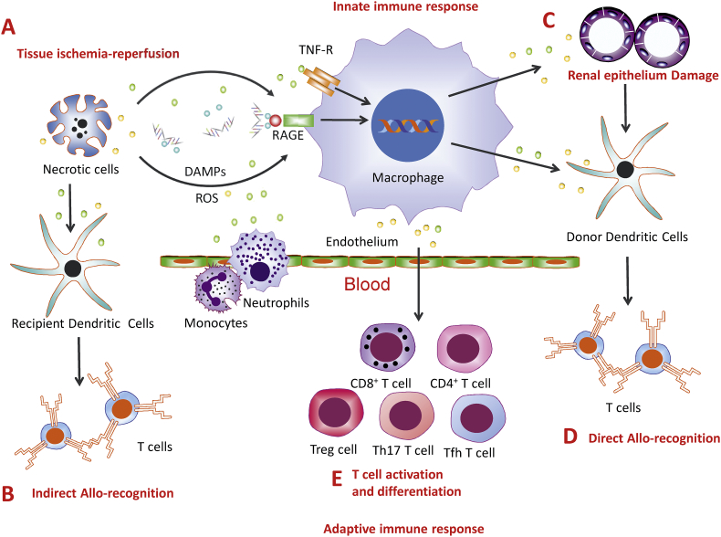
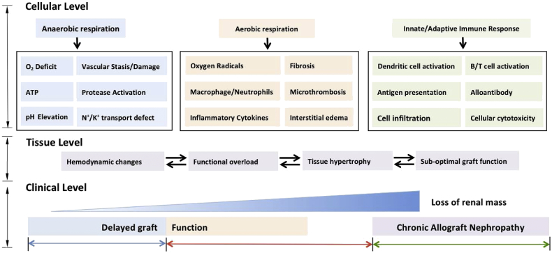
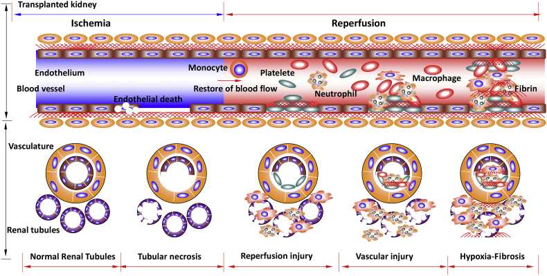
Ischemia-reperfusion injury (IRI) during renal transplantation often initiates non-specific inflammatory responses that can result in the loss of kidney graft viability. However, the long-term consequence of IRI on renal grafts survival is uncertain. Here we review clinical evidence and laboratory studies, and elucidate the association between early IRI and later graft loss. Our critical analysis of previous publications indicates that early IRI does contribute to later graft loss through reduction of renal functional mass, graft vascular injury, and chronic hypoxia, as well as subsequent fibrosis. IRI is also known to induce kidney allograft dysfunction and acute rejection, reducing graft survival. Therefore, attempts have been made to substitute traditional preserving solutions with novel agents, yielding promising results.

Keywords: Acute rejection; Graft survival; Ischemia-reperfusion; Renal transplantation; Th cells: T helper cells.


Figures

Fig. 1
Fig. 1
Ischemia-reperfusion injury and allo-immune response. During renal graft ischemia-reperfusion injury (IRI), (A) renal cell necrosis releases DAMP molecules such as High-mobility group box 1 protein (HMGB1), which are recognised by receptors, such as receptor for advanced glycation end products (RAGE) and Toll-like receptors-2, 4, and 9 (TLR-2, 4 and 9), reactive oxygen species (ROS) and inflammatory cytokines, such as TNF, leading to immune cell activation. (B) Inflammatory cells produce and release pro-inflammatory chemokines (e.g. CXCL1, CXCL2) and cytokines (e.g. IL-1 and 6) and promote tissue inflammation, infiltration of monocyte and neutrophils. (C) Infiltrating monocytes produce ROS and inflammatory cytokines such as TNF, enhance the necroptosis in the epithelial cells (D) Renal cells undergoing necrosis could lead to the release of donor antigens. They are presented by donor antigen presenting cells (APC) and stimulated T-cells. This is direct allo-recognition. (E) As the number of donor antigen presenting cells gradually decreases, the recipient APCs process and present the antigens to T-cells, leading to indirect allo-recognition. (F) The inflammatory milieu formed during IRI influences the activation and differentiation of T-cells, such as CD8+ T-cells, CD4+ Th1 and Th2 cells, Treg cells, Tfh cells and Th17 cells, which contribute to either rejection or tolerance of the renal grafts.
Fig. 2
Fig. 2
Loss of functional mass during renal graft ischemia-reperfusion injury. Renal graft ischemia-reperfusion injury promotes abnormal cellular changes that result in the gradual loss of functional kidney mass. The pathophysiology of ischemia-reperfusion injury is complex and can potentiate graft damage both acutely and chronically due to a variety of cellular and tissue changes that can manifest clinically. On a cellular level, ischemia results in the predominance of anaerobic processes and subsequent cellular damage via processes such as protease activation and N+/K+ transport defects. The associated reperfusion injury occurs due to the restoration of oxygen supply to the graft, resulting in a potpourri of cellular changes including the generation of reactive oxygen species and the upregulation of inflammatory cytokine pathways. The cellular responses during ischemia and reperfusion injury result in abnormal changes within renal tissue, with many of these processes acting synergistically to cause the gradual loss of functional renal mass. The cellular and tissue responses result in graft damage that is exhibited clinically in the form of acute renal necrosis, vasculitis and failure. A less familiar clinical feature of ischemia-reperfusion injury is chronic allograft dysfunction, which predominantly occurs due to abnormal immune activation and the subsequent deterioration in functional graft mass.
Fig. 3
Fig. 3
Graft vascular injury during renal graft ischemia-reperfusion injury. Renal graft vascular injury is a common occurrence following renal transplantation due to ischemia-reperfusion injury. Endothelial vasoconstriction and necrosis occurs during periods of prolonged ischemia. Vasoactive hormones may be responsible for the haemodynamic abnormalities during ischemia, due to an increase in the intrarenal activity of vasoconstrictors and a deficiency of vasodilators. This ultimately results in a reduction in renal blood flow and persistent intrarenal hypoxia, thus exacerbating tubular injury and potentiating acute tubular necrosis within the transplanted kidney. During reperfusion, pro-coagulant pathways predominate, resulting in fibrin deposition and platelet aggregation in peritiubular and glomerular capillaries. These processes can cause microthromboses, leading to impaired renal perfusion and decreased glomerular filtration rate. Red blood cells and leukocytes adhere to the vascular endothelium, further contributing to the blockage of renal blood vessels. The formation of platelet plugs containing fibrin can cause tubular obstruction and increased tubular pressure, it is thought that the exposure of tissue factor to the subendothelium following microvasculature damage promotes the coagulation cascade during reperfusion. Thromboses within the renal vasculature of the transplanted graft promote tubular necrosis and graft failure.

References

    1. Amin R., Turner C., van Aken S., Bahu T.K., Watts A., Lindsell D.R., Dalton R.N., Dunger D.B. The relationship between microalbuminuria and glomerular filtration rate in young type 1 diabetic subjects: the Oxford Regional Prospective Study. Kidney Int. 2005;68:1740–1749. - PubMed
    1. Ascon M., Ascon D.B., Liu M., Cheadle C., Sarkar C., Racusen L., Hassoun H.T., Rabb H. Renal ischemia-reperfusion leads to long term infiltration of activated and effector-memory T lymphocytes. Kidney Int. 2009;75:526–535. - PMC - PubMed
    1. Barba J., Zudaire J.J., Robles J.E., Tienza A., Rosell D., Berian J.M., Pascual I. Is there a safe cold ischemia time interval for the renal graft? Actas Urol. Esp. 2011;35:475–480. - PubMed
    1. Basile D.P., Donohoe D., Roethe K., Osborn J.L. Renal ischemic injury results in permanent damage to peritubular capillaries and influences long-term function. Am. J. Physiol. Renal Physiol. 2001;281:F887–99. - PubMed
    1. Benichou G., Thomson A.W. Direct versus indirect allorecognition pathways: on the right track. Am. J. Transplant. 2009;9:655–656. - PMC - PubMed