. 2018 Feb:28:274-286.
doi: 10.1016/j.ebiom.2018.01.024. Epub 2018 Jan 31.

Allergic Conjunctivitis-induced Retinal Inflammation Promotes Myopia Progression

Affiliations

Allergic Conjunctivitis-induced Retinal Inflammation Promotes Myopia Progression

Chang-Ching Wei et al. EBioMedicine. 2018 Feb.

Erratum in

Abstract

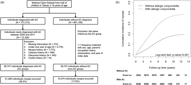
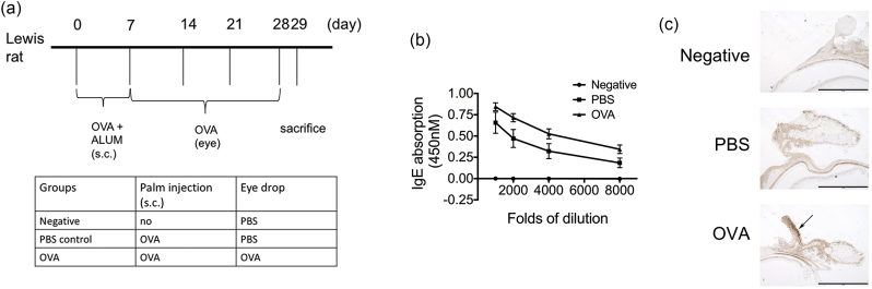
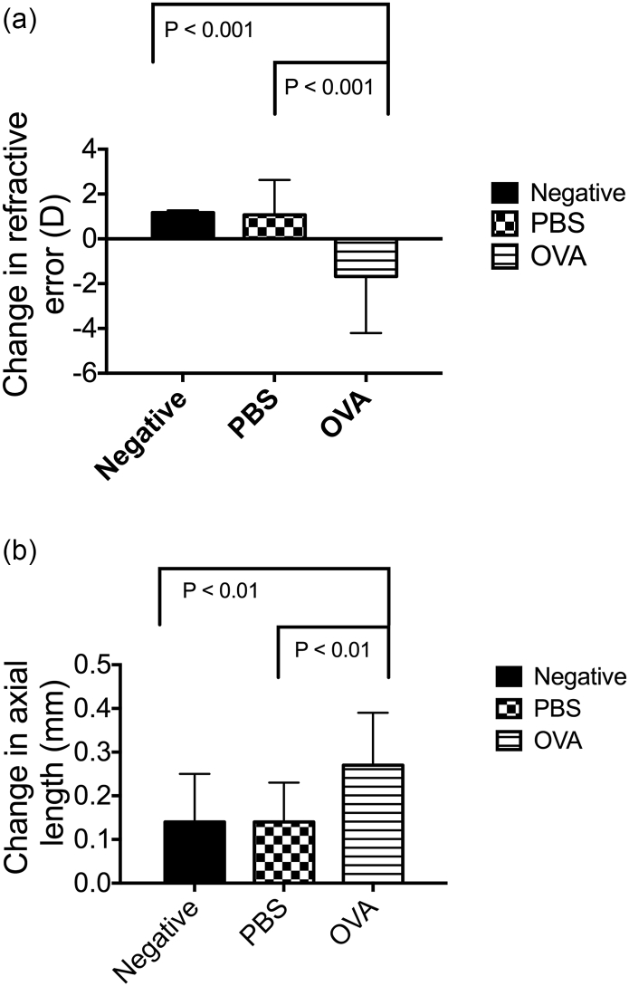
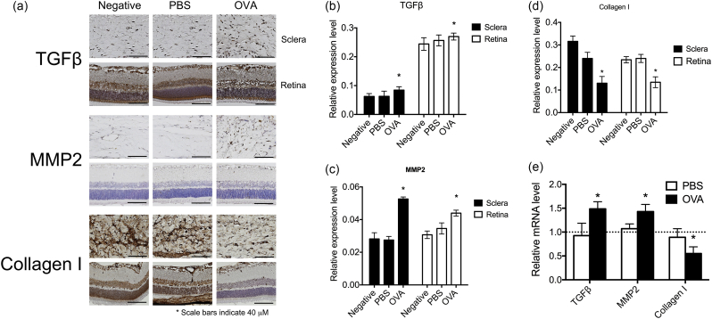
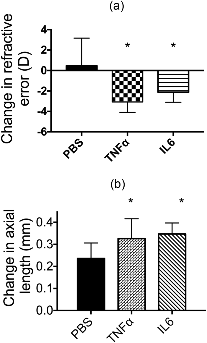
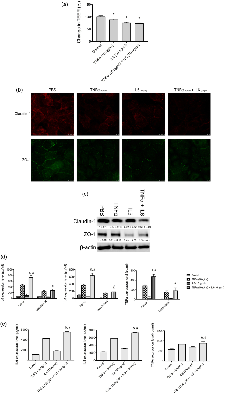
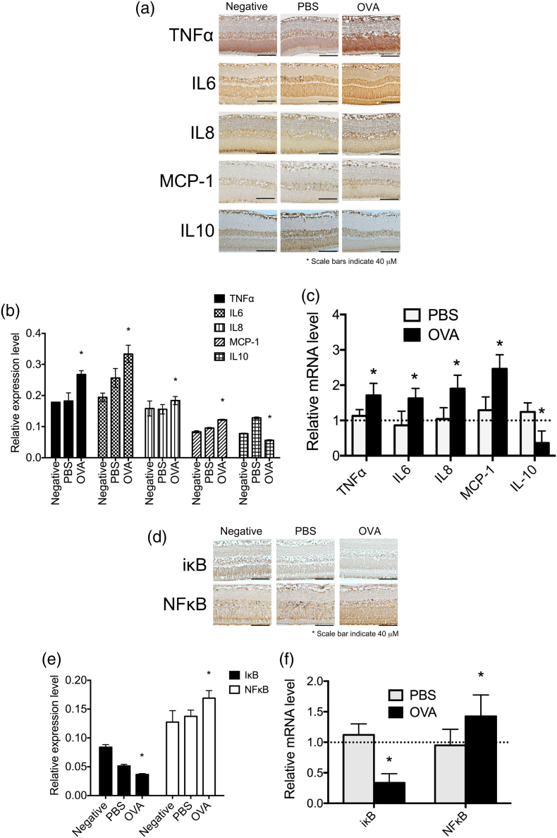
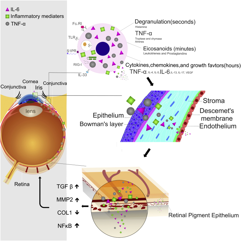
Myopia is a highly prevalent eye disease. There is limited information suggesting a relationship between myopia and inflammation. We found children with allergic conjunctivitis (AC) had the highest adjusted odds ratio (1.75, 95% confidence interval [CI], 1.72-1.77) for myopia among the four allergic diseases. A cohort study was conducted and confirmed that children with AC had a higher incidence and subsequent risk of myopia (hazard ratio 2.35, 95%CI 2.29-2.40) compared to those without AC. Lower refractive error and longer axial length were observed in an AC animal model. Myopia progression was enhanced by tumor necrosis factor (TNF)-α or interleukin (IL)-6 administration, two cytokines secreted by mast cell degranulation. The TNF-α or IL-6 weakened the tight junction formed by corneal epithelial (CEP) cells and inflammatory cytokines across the layer of CEP cells, which increased the levels of TNF-α, IL-6, and IL-8 secreted by retinal pigment epithelial cells. The expression levels of TNF-α, IL-6, IL-8, monocyte chemoattractant protein-1, and nuclear factor kappa B were up-regulated in eyes with AC, whereas IL-10 and the inhibitor of kappa B were down-regulated. In conclusion, the experimental findings in mice corroborate the epidemiological data showing that allergic inflammation influences the development of myopia.

Keywords: Allergic conjunctivitis; Allergic rhinitis; Asthma; Atopic dermatitis; Inflammation; Myopia; Population-based study.


Figures

Fig. 1
Fig. 1
(a) Inclusion and exclusion criteria for the cohort study on allergic conjunctivitis. (b) The association between allergic conjunctivitis (AC) and myopia incidence. The cumulative incidence of myopia is shown for individuals with and without allergic conjunctivitis.
Fig. 2
Fig. 2
Ovalbumin (OVA) administration induced the development of allergic conjunctivitis. (a) The experimental protocol and grouping conditions. (b) Immunoglobulin (Ig)-E antibody secretion in the sera of OVA-sensitized mice. (c) Infiltration of eosinophils (arrow) into the conjunctiva induced by OVA.
Fig. 3
Fig. 3
Allergic conjunctivitis promotes myopia progression. (a) The RE was determined as the difference in diopter measurements taken before and after AC. An ANOVA was used to determine significant differences (P = 0.0011), and Dunnett's multiple comparisons tests were used for paired comparisons between negative, PBS-treated, and OVA-treated eyes. P < 0.05 was considered statistically significant. (b) The axial length was determined as the difference in axial length measurements taken before and after AC. ANOVA was used to determine significant differences (P = 0.0155) and Dunnett's multiple comparisons tests were used for paired comparisons between negative, PBS-treated, and OVA-treated eyes. P < 0.05 was considered statistically significant.
Fig. 4
Fig. 4
Allergic conjunctivitis increased the expression of myopia-related tissue-remodeling proteins. (a) Immunohistochemical analyses of TGF-β, MMP2, and COL-I in negative, PBS-treated, and OVA-treated eyes are shown. (b–d) The relative expression levels of TGF-β (b), MMP2 (c), and COL-I (d) were determined with the Image J software (Schindelin et al., 2012). (e) mRNA expression levels of TGF-β, MMP2, and COL-I in the retinas of PBS and OVA groups were compared to those in the negative group. ANOVA was used to determine significant differences (p < 0.0001) and Sidak's multiple comparisons tests were used for paired comparisons between PBS- and OVA-treated eyes. P < 0.05 was considered statistically significant. *: indicates a significant difference.
Fig. 5
Fig. 5
Effect of increasing inflammation in the eye on myopia progression. (a) The RE was determined as the difference in diopter measurements taken before and after treatment with 10 ng/mL TNF-α or IL-6 for 21 days. ANOVA was used to determine significant differences (p < 0.001), and Tukey's multiple comparisons tests were used for paired comparisons between PBS and TNF-α or IL-6-treated eyes. P < 0.05 was considered statistically significant. (b) The axial length was determined as the difference in axial length measurements taken before and after treatment with 10 ng/mL TNF-α or IL-6 for 21 days. An ANOVA was used to determine significant differences (p = 0.0043), and Tukey's multiple comparisons tests were used for paired comparisons between PBS and TNF-α or IL-6-treated eyes. P < 0.05 was considered statistically significant.
Fig. 6
Fig. 6
TNF-α and IL-6 altered the barrier function of corneal epithelial cells. (a) Relative transepithelial electrical resistance (TEER) of human primary corneal epithelial cells (CEP) incubated with PBS (control), TNF-α (10 ng/mL), IL-6 (10 ng/mL), or TNF-α (10 ng/mL) + IL-6 (10 ng/mL) for 24 h. ANOVA was used to determine significant differences (p < 0.0001), and Dunnett's multiple comparisons tests were used for paired comparisons between PBS and TNF-α or IL-6-treated groups. P < 0.05 was considered statistically significant. (b) CEP cells were cultured in chamber slides at 5 × 104 cells per well and incubated with PBS (control), TNF-α (10 ng/mL), IL-6 (10 ng/mL), or TNF-α (10 ng/mL) + IL-6 (10 ng/mL) for 24 h. Claudin-1 and ZO-1 were detected by immunofluorescence staining. (c) CEP cells were cultured in 6-well plates at 1 × 105 cells per well and incubated with PBS (control), TNF-α (10 ng/mL), IL-6 (10 ng/mL) or TNF-α (10 ng/mL) + IL-6 (10 ng/mL) for 24 h. Claudin-1 and ZO-1 were detected by western blot analysis. The relative expression levels were determined by densitometry analysis. Depicted western blots are one representative figure of three independent experiments. Results are the mean ± S.D. of three independent experiments. (d) CEP cells were seeded on transwell inserts with 0.4-μm pore size incubated with PBS (control), TNF-α (10 ng/mL), IL-6 (10 ng/mL) or TNF-α (10 ng/mL) + IL-6 (10 ng/mL) for 16 h and the levels of TNF-α, IL-6, and IL-8 in apical and basolateral compartments were determined with enzyme-linked immunosorbent assay (ELISA). ANOVA was used to determine significant differences (p < 0.0001), and Tukey's multiple comparisons tests were used for paired comparisons between TNF-α (&) or IL-6 (#) with TNF-α + IL-6-treated CEP cells. P < 0.05 was considered statistically significant. (e) The basolateral compartment media of CEP cells treated with PBS (control), TNF-α (10 ng/mL), IL-6 (10 ng/mL), or TNF-α (10 ng/mL) + IL-6 (10 ng/mL) for 16 h were collected. The basolateral compartment media were used to treat human primary retinal pigment epithelial cells for 16 h, and the levels of TNF-α, IL-6, and IL-8 were determined with ELISA. ANOVA was used to determine significant differences (p < 0.0001), and Tukey's multiple comparisons tests were used for paired comparisons between TNF-α (&) or IL-6 (#) with TNF-α + IL-6-treated retinal pigment epithelial cells. P < 0.05 was considered statistically significant.
Fig. 7
Fig. 7
Expression levels of inflammation-related transcription factors and cytokines in allergic conjunctivitis eyes. (a) Immunohistochemical analyses of TNF-α, IL-6, IL-8, MCP-1, and IL-10 expressions in negative, PBS-treated, and OVA-treated eyes. (b) The relative expression levels of TNF-α, IL-6, IL-8, MCP-1, and IL-10 were determined with the Image J software. (c) mRNA expression levels of TNF-α, IL-6, IL-8, MCP-1, and IL-10 in the retinas of the PBS and OVA groups were compared to those in the negative group. ANOVA was used to determine significant differences (p < 0.0001) and Sidak's multiple comparisons test was used for paired comparisons between PBS- and OVA-treated eyes. P < 0.05 was considered statistically significant. *: indicates a significant difference. (d) Immunohistochemical analyses of NFκB and IκB expressions in negative, PBS-treated, and OVA-treated eyes. (e) The relative expression levels of NFκB and IκB were determined with the Image J software. (f) mRNA expression levels of NFκB and IκB in the retinas of the PBS and OVA groups were compared to those in the negative group. ANOVA was used to determine significant differences (p < 0.0001) and Sidak's multiple comparisons test was used for paired comparisons between PBS- and OVA-treated eyes. P < 0.05 was considered statistically significant. *: indicates a significant difference.
Fig. 8
Fig. 8
Schematic presentation of how allergic conjunctivitis inflammation modulates the signaling pathway to promote myopia.

Similar articles

Cited by

References

    1. Abraham E., Arcaroli J., Shenkar R. Activation of extracellular signal-regulated kinases, NF-kappa B, and cyclic adenosine 5′-monophosphate response element-binding protein in lung neutrophils occurs by differing mechanisms after hemorrhage or endotoxemia. J. Immunol. 2001;166(1):522–530. - PubMed
    1. Akasu T., Tsurusaki M. Interleukin-1beta causes a biphasic response in neurons of rat major pelvic ganglia. Neurosci. Lett. 1999;272(2):119–122. - PubMed
    1. Bacic F., Uematsu S., McCarron R.M., Spatz M. Dopaminergic receptors linked to adenylate cyclase in human cerebromicrovascular endothelium. J. Neurochem. 1991;57(5):1774–1780. - PubMed
    1. Basu S., Dasgupta P.S. Dopamine, a neurotransmitter, influences the immune system. J. Neuroimmunol. 2000;102(2):113–124. - PubMed
    1. Beck G., Brinkkoetter P., Hanusch C. Clinical review: immunomodulatory effects of dopamine in general inflammation. Crit. Care. 2004;8(6):485–491. - PMC - PubMed

MeSH terms