. 2018 Feb 14;23(2):203-214.e5.
doi: 10.1016/j.chom.2018.01.007. Epub 2018 Feb 1.

Bacteroides fragilis Toxin Coordinates a Pro-carcinogenic Inflammatory Cascade via Targeting of Colonic Epithelial Cells

Affiliations

Bacteroides fragilis Toxin Coordinates a Pro-carcinogenic Inflammatory Cascade via Targeting of Colonic Epithelial Cells

Liam Chung et al. Cell Host Microbe. .

Erratum in

Abstract

Pro-carcinogenic bacteria have the potential to initiate and/or promote colon cancer, in part via immune mechanisms that are incompletely understood. Using ApcMin mice colonized with the human pathobiont enterotoxigenic Bacteroides fragilis (ETBF) as a model of microbe-induced colon tumorigenesis, we show that the Bacteroides fragilis toxin (BFT) triggers a pro-carcinogenic, multi-step inflammatory cascade requiring IL-17R, NF-κB, and Stat3 signaling in colonic epithelial cells (CECs). Although necessary, Stat3 activation in CECs is not sufficient to trigger ETBF colon tumorigenesis. Notably, IL-17-dependent NF-κB activation in CECs induces a proximal to distal mucosal gradient of C-X-C chemokines, including CXCL1, that mediates the recruitment of CXCR2-expressing polymorphonuclear immature myeloid cells with parallel onset of ETBF-mediated distal colon tumorigenesis. Thus, BFT induces a pro-carcinogenic signaling relay from the CEC to a mucosal Th17 response that results in selective NF-κB activation in distal colon CECs, which collectively triggers myeloid-cell-dependent distal colon tumorigenesis.

Keywords: Nf-κB; STAT-3; colorectal cancer; inflammation; mucosal immunology; myeloid cells.


Conflict of interest statement

DECLARATION OF INTERESTS

The authors declare no competing interests

Figures

Figure 1
Figure 1. BFT and IL-17 target colonic epithelial cells to promote ETBF-mediated carcinogenesis
A, colon tumor numbers in ApcMin mice colonized with ETBF or ETBF(Δbft) for a period of 8 weeks. Non-colonized ApcMin mice (Sham) were used as control. B, colon tumor numbers in parental (Il17a+/+Il17ra+/+), IL-17A deficient (Il17a−/−Il17ra+/+) and IL-17RA deficient (Il17a+/+Il17ra−/−) ApcMin mice 12 weeks after ETBF colonization. C, colon tumor numbers in [WT(IL17ra+/+)> ApcMin] and [Il17a−/− C57BL/6 > ApcMin] BM chimera mice 12 weeks after ETBF colonization. D, colon tumor numbers in [Il17a+/+(WT)>ApcMin] and [WT>Il17ra−/− ApcMin] BM chimera mice 12 weeks after ETBF colonization. Graphs show mean+/− SEM. E, Immunofluorescent microscopic analysis of IL-17RA expression in 4 week ETBF-colonized ApcMin mice. Blue, DAPI; green, E-cadherin (epithelial cells); yellow, SMA (fibroblasts and myofibroblasts); red, IL-17RA. Dotted line delineates microadenoma. Scale bar, 50 μm
Figure 2
Figure 2. IL-17 dependent NF-κb signaling in CECs triggers higher expression of Cxcl1, Cxcl2 and Cxcl5 in the distal compared to proximal segments of ETBF-colonized colon
A, Il17a, Il17ra, Il17rc, Cxcl1, Cxcl2 and Cxcl5 mRNA expression in whole tissue sections from proximal (P; C6) to distal (D; C1) colon of WT (Sham, blue circles; ETBF, red squares), Il17a−/− (Il17a−/−, ETBF, red triangles) and Il17ra−/− (Il17ra−/−, ETBF, red stars) C57BL/6 (WT) mice. A representative image of colon segments C6 to C1 is shown. In panels B–D these segments are grouped into P (C6, C5), M (mid, C4, C3) and D (C2, C1). Ct are normalized with gapdh Ct (ΔCt) and mRNA expression is calculated as 1000x2−ΔCt. B, CECs were isolated from proximal (P), mid (M) and distal (D) portions of colons obtained from either Sham or ETBF-colonized C57BL/6 mice at day 7 post infection. Top, nuclear fractions derived from the indicated CECs were immunoblotted (IB) for p65 and pStat3. Caspase-3 (Casp3) and PARP1 served as loading controls for cytosolic and nuclear markers, respectively. Bottom, whole cell lysates (WCL) derived from the indicated CECs were IB for IL-17R, with β-actin as a loading control. Data are representative of two independent experiments. C, CECs were isolated from the mid (M) and distal (D) portions of colons obtained from either Sham or ETBF-colonized IL-17ra+/+ (WT) or IL-17ra−/− C57BL/6 mice at day 7 post-infection. D, HT29/c1 cells were stimulated with the indicated concentration of TNFα, purified BFT or IL-17a for 6h. Nuclear fractions were derived and IB for p65. Casp3 and PARP1 served as loading controls and cytosolic and nuclear markers, respectively.
Figure 3
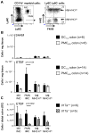
Figure 3. IL-17-dependent accumulation of IMCs in the distal part of the colon upon ETBF colonization of WT mice
A, Gating strategy used to delineate the myeloid population in enzymatically digested colonic LP 7 days after ETBF colonization of WT C57BL/6 mice. MO-IMC and PMN-IMC are defined as CD45+CD3CD11bhiLy6ChiLy6G and CD45+CD3CD11bhiLy6CintLy6G+ respectively. Macrophages (Mϕ) are characterized as CD45+CD3CD11bhiLy6CLy6GF4/80+I-A/Ehi (Mϕ-MHC-IIhi) and CD45+CD3CD11bhiLy6C−iLy6GF4/80+I-A/Elo (Mϕ-MHC-IIlo). B, Numbers of myeloid cells per mg of tissues in the distal (DC1–2, C1–C2) (black bars) and mid-proximal (PMC4–5, sections C4–C5, Figure 2) (open bars) colon isolated from Sham C57BL/6 mice (top graph; n=8) and 7 days after ETBF colonization (bottom panel; n=14). % of alive CD45 cells. C, Number of myeloid cells in the distal colon (DC) colons isolated from WT (Il17a+/+; black bars; n=6) and IL17a−/− C57BL/6 mice (striped bars; n=5) 7 days after ETBF colonization. % of alive CD45 cells shown. Graphs show mean+/− SEM.
Figure 4
Figure 4. Myeloid recruitment and colon tumorigenesis are impaired upon Cxcr2 deletion or pepducin-mediated inhibition of CXCR2 in ETBF-colonized ApcMin mice
A, Myeloid cell recruitment in the distal colon (DC) of CXCr2+/+ WT (black bars; n=8) and CXCR2−/− (open bars; n=7) mice 7 days after ETBF colonization. MO-IMC, CD11b+Ly6ChiLy6GF4/80I-A/Elo; PMN-IMC, CD11b+Ly6CloLy6G+F4/80I-A/E-; Mϕ-MHC-IIhi, CD11b+Ly6CLy6GF4/80+I-A/Ehi; Mϕ-MHC-IIlo, CD11b+Ly6CLy6GF4/80+I-A/Elo. B, Number of IFNγ- and IL-17-producing cells in the distal colon (DC) of CXCr2+/+ WT (black bars; n=8) and CXCR2−/− (white bars; n=7) mice 7 days after ETBF colonization. C, colon tumor numbers in control [CXCr2+/+>ApcMin] (n=10) and [CXCr2−/−>ApcMin] (n=10) BM chimera mice 12 weeks after ETBF colonization. D, Myeloid cell recruitment in the distal colon (DC) of WT mice treated with control peptide (ctrl) (black bars; n=4) and the CXCR2 inhibitor pepducin (open bars; n=7) mice 7 days after ETBF colonization. Graphs show mean+/− SEM. E, Il17a, Cxcl1, Cxcl2 and Cxcl5 gene expression in whole colon tissue (DC1–2 portion) of ctrl peptide (black symbols) and the CXCR2 inhibitor pepducin (open symbols)-treated WT mice 7 days after ETBF colonization. Ct are normalized with gapdh Ct (ΔCt) and mRNA expression is calculated as 1000x2−ΔCt. Graphs show mean+/− SEM. F, microadenoma numbers in ApcMin mice treated with ctrl peptide and the CXCR2 inhibitor pepducin 4 weeks after ETBF colonization. Graphs show mean+/− SEM. A representative micrograph of a colon microadenoma is shown in insert. Scale bar, 50 μm.
Figure 5
Figure 5. ETBF-triggered colon tumorigenesis in ApcMin mice is decreased in absence of epithelial Stat3 signaling
A, colon tumor numbers in Stat3+/+ parental (black square; n=8) and Stat3Δ IEC(open squares; n=18) ApcMin mice 12 weeks after ETBF colonization. B, myeloid cell recruitment in the distal colon (DC) of Stat3+/+ WT (black bars; n=6) and Stat3Δ IEC (open bars; n=5) mice 7 days after ETBF colonization. MO-IMC, CD11b+Ly6ChiLy6GF4/80I-A/Elo; PMN-IMC, CD11b+Ly6CloLy6G+F4/80I-A/E-; Mϕ-MHC-IIhi, CD11b+Ly6CLy6G F4/80+ I-A/Ehi; Mϕ-MHC-IIlo, CD11b+Ly6CLy6G F4/80+ I-A/Elo. C, Number of IFNγ– and IL-17-producing cells in the distal (DC) colon of Stat3+/+ WT (black bars; n=6) and Stat3Δ IEC (white bars; n=5) mice 7 days after ETBF colonization. Graphs show mean+/− SEM. D, Il17a gene expression in whole colon tissue (DC1–2) of IECStat3+/+ WT (n=4) and IECStat3−/− (n=6) mice 7 days after ETBF colonization. Ct are normalized with gapdh Ct (ΔCt). Il17a expression is indicated as ratio (RQ) of ΔCt in ETBF mice to ΔCt in Sham mice (ET/Sham). Graphs show mean+/− SEM. E, colon tumor numbers in Il6+/+ parental (Sham, black circle; n=3 and ETBF, black square;n=7) and Il6−/− (Sham, open circle; n=4 and ETBF, open square; n=12) ApcMin mice 12 weeks after ETBF colonization. Graphs show mean+/− SEM. F, Il17a gene expression in whole colon tissue of WT (open circle, D3; open square, D7) and IL6−/− (black circle, D3; black square, D7) mice 3 and 7 days after ETBF colonization. Ct are normalized with gapdh Ct (ΔCt) and mRNA expression is calculated as 1000x2−ΔCt. Graphs show mean+/− SEM.
Figure 6
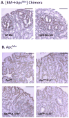
Figure 6. Epithelial Stat3 remains activated in ETBF-colonized ApcMin mice in absence of IL-17, IL-6 or CXCR2 expression
A, pStat3 IHC in [CXCr2+/+>ApcMin] (WT BM; left), [CXCr2−/−>ApcMin] (CXCR2KO BM; right); B, parental ApcMin (upper left), Il6−/−ApcMin (upper right), Il17ra−/−ApcMin (bottom left) and Il22−/− ApcMin. All mice were sacrificed 12 weeks after ETBF colonization. scale bars, 100um.
Figure 7
Figure 7. Schematic representation of the mechanistic CEC:mucosal relay promoting ETBF-induced colon tumorigenesis
Collectively the data herein support a model whereby ETBF infection triggers a specific relay of cellular interactions initiated by the action of the single bacterial virulence protein, BFT, on CECs that results first in IL-17-producing mucosal immune cells that then turn to act on the CECs to initiate a NFκB-associated CEC immune response that directs distal colon myeloid mucosal infiltration key to ETBF tumor localization in the distal colon. ETBF orchestrated colon carcinogenesis occurs in the absence of IL-6 and IL-23. CEC Stat3 activation is a necessary, but not sufficient, complement to the actions of IL-17. Steps in this model include: 1, ETBF produces BFT, a zinc-metalloprotease that induces E-cadherin cleavage that contributes to barrier permeability dysfunction as well as Wnt/βcatenin and NF-κB signaling pathway activation. 2, a diminished colonic mucosal barrier likely contributes to resident myeloid cells activation that contributes to the rapid onset ETBF IL-17 signature characterized by the accumulation of Th17, γδT17 and ILC3. Further, dependent on redundant cytokines due to the BFT virulence protein of ETBF, Stat3 is highly activated in lamina propia ICs including lymphocytes and myeloid cells. 3, IL-17/IL-17R and BFT signaling pathways converge on CECs to activate a distal colon NF-κB cascade resulting in the production of a C-X-C chemokine distal colon mucosal gradient, including CXCL1 (homologous to human IL-8). 4, although the ETBF-altered colon barrier progressively recovers its integrity, ETBF persistence maintains IL-17 production and the chemokine gradient that promotes the accumulation of distal colon MDSCs, including high proportions of PMN-MDSC. 5, the oncogenic properties of BFT including BFT-induced CEC proliferation and DNA damage likely allow the rapid loss of the second Apc allele (loss of heterozygosity, LOH) and rapid promotion of microadenoma formation (DeStefano Shields et al., 2016; Wu et al., 2003). MDSC accumulation in the distal colon provides growth factors promoting adenoma growth.

Comment in

References

    1. Acharyya S, Oskarsson T, Vanharanta S, Malladi S, Kim J, Morris PG, Manova-Todorova K, Leversha M, Hogg N, Seshan VE, et al. A CXCL1 paracrine network links cancer chemoresistance and metastasis. Cell. 2012;150:165–178. - PMC - PubMed
    1. Boleij A, Hechenbleikner EM, Goodwin AC, Badani R, Stein EM, Lazarev MG, Ellis B, Carroll KC, Albesiano E, Wick EC, et al. The Bacteroides fragilis toxin gene is prevalent in the colon mucosa of colorectal cancer patients. Clin Infect Dis. 2015;60:208–215. - PMC - PubMed
    1. Bollrath J, Phesse TJ, von Burstin VA, Putoczki T, Bennecke M, Bateman T, Nebelsiek T, Lundgren-May T, Canli O, Schwitalla S, et al. gp130-mediated Stat3 activation in enterocytes regulates cell survival and cell-cycle progression during colitis-associated tumorigenesis. Cancer cell. 2009;15:91–102. - PubMed
    1. Chen K, Eddens T, Trevejo-Nunez G, Way EE, Elsegeiny W, Ricks DM, Garg AV, Erb CJ, Bo M, Wang T, et al. IL-17 Receptor Signaling in the Lung Epithelium Is Required for Mucosal Chemokine Gradients and Pulmonary Host Defense against K. pneumoniae. Cell Host Microbe. 2016;20:596–605. - PMC - PubMed
    1. Conti HR, Bruno VM, Childs EE, Daugherty S, Hunter JP, Mengesha BG, Saevig DL, Hendricks MR, Coleman BM, Brane L, et al. IL-17 Receptor Signaling in Oral Epithelial Cells Is Critical for Protection against Oropharyngeal Candidiasis. Cell Host Microbe. 2016;20:606–617. - PMC - PubMed

MeSH terms