Review
. 2018 Jun;32(6):1010-1020.
doi: 10.1038/s41433-018-0021-7. Epub 2018 Feb 5.

Clinical pharmacology of intravitreal anti-VEGF drugs

Affiliations
Review

Clinical pharmacology of intravitreal anti-VEGF drugs

Stefano Fogli et al. Eye (Lond). 2018 Jun.

Abstract

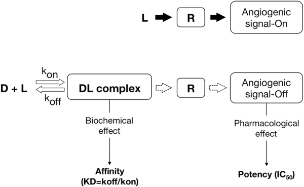
Clinical efficacy of intravitreal anti-VEGF drugs has been widely demonstrated in several angiogenesis-driven eye diseases including diabetic macular edema and the neovascular form of age-related macular degeneration. Pegaptanib, ranibizumab, and aflibercept have been approved for use in the eye, whereas bevacizumab is widely used by ophthalmologists to treat patients "off-label". These drugs are active in the nanomolar to picomolar range; however, caution is required when establishing the rank order of affinity and potency due to in vitro inter-experimental variation. Despite the small doses used for eye diseases and the intravitreal route of administration may limit systemic side effects, these drugs can penetrate into blood circulation and alter systemic VEGF with unknown clinical consequences, particularly in vulnerable groups of patients. Clinical pharmacokinetics of ocular anti-VEGF agents should therefore be taken into account when choosing the right drug for the individual patient. The gaps in current understanding that leave open important questions are as follows: (i) uncertainty about which drug should be given first, (ii) how long these drugs can be used safely, and (iii) the choice of the best pharmacological strategy after first-line treatment failure. The current review article, based on the information published in peer-reviewed published papers relevant to anti-VEGF treatments and available on the PubMed database, describes in detail the clinical pharmacology of this class of drugs to provide a sound pharmacological basis for their proper use in ophthalmology clinical practice.


Conflict of interest statement

The authors declare that they have no competing interests.

Figures

Fig. 1
Fig. 1
Mechanism of action of intravitreal antiangiogenic drugs. Angiogenic factors (e.g., VEGF) stimulate their specific receptors (e.g., VEGFR) activating the signaling cascade. Binding of intravitreal antiangiogenic drugs to their ligands (e.g., VEGF) form the drug–ligand complex (DL) preventing ligand/receptor interaction and keeping (or turning) the angiogenic switch off. Affinity is defined as the degree of attraction between drug and the target, and it is expressed by the dissociation constant, Kd (that is, the ratio between dissociation (Koff) and association (Kon) rates). Kd is the inverse of the affinity to the binding site (i.e., the lower the Kd, the higher the affinity). Potency is the amount of drug needed to produce the pharmacological effect. For these drugs, potency is expressed by the half maximal inhibitory concentration (IC50), a measure of the effectiveness of a substance in inhibiting a specific biological function (the lower the IC50, the higher the potency)
Fig. 2
Fig. 2
Pharmacokinetic model for intravitreal antiangiogenic drugs. Intravitreally injected Fc-carrying molecules can penetrate into the systemic circulation by a FcRn-dependent transport. The presence of the Fc portion may also reduce systemic clearance thus prolonging drug exposure (see text for further details). Renal clearance may depend on the molecular size; in particular, the higher the molecular weight, the lower the drug clearance (e.g., pegylated molecules, full-length monoclonal antibodies). FcRn neonatal Fc receptor, BRB blood retinal barrier

References

    1. Fogli S, Mogavero S, Egan CG, Del Re M, Danesi R. Pathophysiology and pharmacological targets of VEGF in diabetic macular edema. Pharmacol Res. 2016;103:149–57. doi: 10.1016/j.phrs.2015.11.003. - DOI - PubMed
    1. Campochiaro PA. Molecular pathogenesis of retinal and choroidal vascular diseases. Prog Retin Eye Res. 2015;49:67–81. doi: 10.1016/j.preteyeres.2015.06.002. - DOI - PMC - PubMed
    1. Senger DR, Davis GE. Angiogenesis. Cold Spring Harb Perspect Biol. 2011;3(8):a005090–a005090. doi: 10.1101/cshperspect.a005090. - DOI - PMC - PubMed
    1. Chalam KV, Brar VS, Murthy RK. Human ciliary epithelium as a source of synthesis and secretion of vascular endothelial growth factor in neovascular glaucoma. JAMA Ophthalmol. 2014;132(11):1350–4. doi: 10.1001/jamaophthalmol.2014.2356. - DOI - PubMed
    1. Kaur C, Foulds WS, Ling EA. Hypoxia-ischemia and retinal ganglion cell damage. Clin Ophthalmol. 2008;2(4):879–89. doi: 10.2147/OPTH.S3361. - DOI - PMC - PubMed

MeSH terms

Substances