. 2018 Jan;15(1):998-1006.
doi: 10.3892/ol.2017.7386. Epub 2017 Nov 9.

Microglia immunophenotyping in gliomas

Affiliations

Microglia immunophenotyping in gliomas

Laura Annovazzi et al. Oncol Lett. 2018 Jan.

Abstract

Microglia, once assimilated to peripheral macrophages, in gliomas has long been discussed and currently it is hypothesized to play a pro-tumor role in tumor progression. Uncertain between M1 and M2 polarization, it exchanges signals with glioma cells to create an immunosuppressive microenvironment and stimulates cell proliferation and migration. Four antibodies are currently used for microglia/macrophage identification in tissues that exhibit different cell forms and cell localization. The aim of the present work was to describe the distribution of the different cell forms and to deduce their significance on the basis of what is known on their function from the literature. Normal resting microglia, reactive microglia, intermediate and bumpy forms and macrophage-like cells can be distinguished by Iba1, CD68, CD16 and CD163 and further categorized by CD11b, CD45, c-MAF and CD98. The number of microglia/macrophages strongly increased from normal cortex and white matter to infiltrating and solid tumors. The ramified microglia accumulated in infiltration areas of both high- and low-grade gliomas, when hypertrophy and hyperplasia occur. In solid tumors, intermediate and bumpy forms prevailed and there is a large increase of macrophage-like cells in glioblastoma. The total number of microglia cells did not vary among the three grades of malignancy, but macrophage-like cells definitely prevailed in high-grade gliomas and frequently expressed CD45 and c-MAF. CD98+ cells were present. Microglia favors tumor progression, but many aspects suggest that the phagocytosing function is maintained. CD98+ cells can be the product of fusion, but also of phagocytosis. Microglia correlated with poorer survival in glioblastoma, when considering CD163+ cells, whereas it did not change prognosis in isocitrate dehydrogenase-mutant low grade gliomas.

Keywords: gliomas; immunosuppression; macrophages; microglia; phagocytosis.


Figures
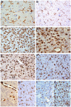
Figure 1.
Figure 1.
Immunohistochemical characterization of microglia/macrophages. (A) Resident ramified microglia in the normal cortex, Iba1 staining, ×400. (B) Diffuse astrocytoma. Reactive ramified microglia in mild tumor infiltration, Iba1 staining, ×200. (C) Diffuse astrocytoma. Reactive ramified microglia in high tumor infiltration, Iba1 staining, ×200. (D) Diffuse astrocytoma. Intermediate and macrophagic forms, CD16 staining, ×200. (E) Oligodendroglioma. Cells with a macrophage-like aspect in solid tumor, CD68 staining, ×200. (F) Oligodendroglioma. Reactive ramified microglia and macrophagic forms, Iba1 staining, ×200. (G) Oligodendroglioma. CD163+ cells were mainly found around vessels, CD163 staining, ×200. (H) Oligodendroglioma. In MIA and HIA Iba1+ RM did not crowd around small vessels, Iba1 staining, ×200. (I) Anaplastic oligodendroglioma. Intermediate and macrophagic forms in solid tumor, Iba1 staining, ×200. (J) Anaplastic astrocytoma. Intermediate and macrophagic forms in solid tumor, CD163 staining, ×200. All stained with DAB. Iba1, allograft inflammatory factor 1; DAB, 3,3′-diaminobenzidine; CD, cluster of differentiation; MIA, mild infiltration area; HIA, high infiltration area; RM, reactive ramified microglia.
Figure 2.
Figure 2.
Immunohistochemical characterization of microglia/macrophages. (A) GB; High frequency of CD16+ intermediate forms in solid tumor, CD16 staining, ×200, DAB. (B) GB; Perivascular CD163+ macrophagic forms, CD163 staining, ×200, DAB. (C) GB; Iba1+ macrophagic forms, Iba1 staining, ×200, DAB. (D) GB; Passage from a solid tumor to a regressive area: CD163+ cells are rare in the former and strongly positive and frequent as macrophagic forms in the latter, CD163 staining, ×200, DAB. (E) GB; Crowding of GAMs in glomerular structures, Iba1 (DAB) and CD34 (RED) stainings, ×200. (F) GB; Macrophagic forms with c-MAF+ nuclei and CD163+ cytoplasms, c-MAF (DAB) and CD163 (RED) stainings, ×630. (G) GB; Perivascular CD45+ M forms, CD45 staining, ×200, DAB. (H) GB; Perivascular CD163+ M forms, CD163 staining, ×200, DAB. (I) GB; CD98+ macrophage-like cells, CD98 staining, ×400, DAB. CD, cluster of differentiation; DAB, 3,3′-diaminobenzidine; HPF, high power fields; GB, Glioblastoma; Iba1, allograft inflammatory factor 1; GAMs, glioma-associated microglia/macrophages.
Figure 3.
Figure 3.
Correlation of the microglia/macrophages markers with the histological grade in 95 patients with WHO II–IV grade gliomas. The number of MM immuno-positive for CD68 and CD163 correlated with the grade of malignancy. Data are mean values ± SE. *P<0.05, one-way ANOVA and Tukey's test. CD, cluster of differentiation; Iba1, allograft inflammatory factor 1; HPF, high power fields.
Figure 4.
Figure 4.
Kaplan-Meier survival analysis in 95 patients of WHO II–IV grade gliomas, according to the CD163 immunohistochemical expression. (A) Correlation of OS with CD163 levels according to a four-category (0, 1, 2, 3) scoring system. (B) Correlation of OS with CD163 levels according to a two-category (low and high) scoring system. The log-rank test was performed to determine statistical significance, with P<0.05 considered as significant. OS, overall survival; CD, cluster of differentiation; WHO, World Health Organization.
Figure 5.
Figure 5.
Kaplan-Meier survival analysis according to the CD163 immunohistochemical expression. (A) Correlation of overall survival (OS) with CD163 levels according to a four-category (0, 1, 2, 3) scoring system in WHO III grade glioma patients. (B) Correlation of OS with the IDH gene status and the CD163 expression (0=low, 1=high) in 60 patients of WHO II–III grade gliomas. The log-rank test was performed to determine statistical significance, and P<0.05 was considered significant. CD, luster of differentiation; M, mutated; WT, wildtype; WHO, World Health Organization; IDH, isocitrate dehydrogenase.

References

    1. Graeber MB, Streit WJ. Microglia: Biology and pathology. Acta Neuropathol. 2010;119:89–105. doi: 10.1007/s00401-009-0622-0. - DOI - PubMed
    1. Lawson LJ, Perry VH, Dri P, Gordon S. Heterogeneity in the distribution and morphology of microglia in the normal adult mouse brain. Neuroscience. 1990;39:151–170. doi: 10.1016/0306-4522(90)90229-W. - DOI - PubMed
    1. Kettenmann H, Hanisch UK, Noda M, Verkhratsky A. Physiology of microglia. Physiol Rev. 2011;91:461–553. doi: 10.1152/physrev.00011.2010. - DOI - PubMed
    1. Simmons GW, Pong WW, Emnett RJ, White CR, Gianino SM, Rodriguez FJ, Gutmann DH. Neurofibromatosis-1 heterozygosity increases microglia in a spatially-and temporally-restricted pattern relevant to mouse optic glioma formation and growth. J Neuropathol Exp Neurol. 2011;70:51–62. doi: 10.1097/NEN.0b013e3182032d37. - DOI - PMC - PubMed
    1. Klein R, Roggendorf W. Increased microglia proliferation separates pilocytic astrocytomas from diffuse astrocytomas: A double labeling study. Acta Neuropathol. 2001;101:245–248. - PubMed