

Contrast uptake in primary hepatic angiosarcoma on gadolinium-ethoxybenzyl-diethylenetriamine pentaacetic acid-enhanced magnetic resonance imaging in the hepatobiliary phase
- PMID: 29399290
- PMCID: PMC5787680
- DOI: 10.4254/wjh.v10.i1.166
Contrast uptake in primary hepatic angiosarcoma on gadolinium-ethoxybenzyl-diethylenetriamine pentaacetic acid-enhanced magnetic resonance imaging in the hepatobiliary phase
Abstract
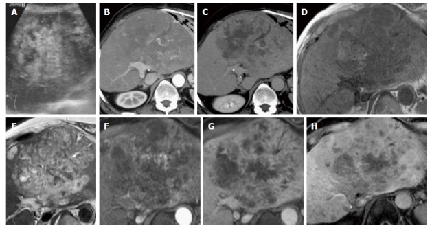
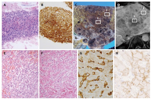
Primary hepatic angiosarcoma is the most common malignant mesenchymal tumor of the liver. It has a poor prognosis and various appearances on magnetic resonance (MR) images. We report a case of hepatic angiosarcoma with a characteristic appearance on gadolinium-ethoxybenzyl-diethylenetriamine pentaacetic acid (Gd-EOB-DTPA)-enhanced MR imaging in the hepatobiliary phase. A 72-year-old man was admitted with a complaint of abdominal pain. Gd-EOB-DTPA-enhanced MR imaging revealed a liver tumor that showed slight hyperintensity in the hepatobiliary phase. These findings suggested Gd-EOB-DTPA uptake in the tumor. An autopsy revealed the solid proliferation and sinusoidal spreading of hepatic angiosarcoma cells. Immunohistochemistry indicated that the tumor was negative for OATP1B3. Gd-EOB-DTPA uptake in the liver tumor in the hepatobiliary phase suggested sinusoidal tumor invasion with residual normal hepatocytes.
Keywords: Cirrhosis; Gadolinium-ethoxybenzyl-diethylenetriamine pentaacetic acid; Hepatic angiosarcoma; Hepatocellular carcinoma.
Conflict of interest statement
Conflict-of-interest statement: There are no conflicts of interest to declare.
Figures
References
-
- Alrenga DP. Primary angiosarcoma of the liver. Review article. Int Surg. 1975;60:198–203. - PubMed
-
- Ishak K, Peters R, editors . Mesenchymal tumor of the liver. Hepatocellular carcinoma. New York: Wiley; 1976. pp. 247–308.
-
- Locker GY, Doroshow JH, Zwelling LA, Chabner BA. The clinical features of hepatic angiosarcoma: a report of four cases and a review of the English literature. Medicine (Baltimore) 1979;58:48–64. - PubMed
-
- Buetow PC, Buck JL, Ros PR, Goodman ZD. Malignant vascular tumors of the liver: radiologic-pathologic correlation. Radiographics. 1994;14:153–166; quiz 167-168. - PubMed
-
- Pickhardt PJ, Kitchin D, Lubner MG, Ganeshan DM, Bhalla S, Covey AM. Primary hepatic angiosarcoma: multi-institutional comprehensive cancer centre review of multiphasic CT and MR imaging in 35 patients. Eur Radiol. 2015;25:315–322. - PubMed
Publication types
LinkOut - more resources
Full Text Sources
Other Literature Sources
