

Proximal Humerus Fractures: Evaluation and Management in the Elderly Patient
- PMID: 29399372
- PMCID: PMC5788098
- DOI: 10.1177/2151458517750516
Proximal Humerus Fractures: Evaluation and Management in the Elderly Patient
Abstract
Introduction: Proximal humerus fractures are common in the elderly. The evaluation and management of these injuries is often controversial. The purpose of this study is to review recent evidence and provide updated recommendations for treating proximal humerus fractures in the elderly.
Methods: A literature review of peer-reviewed publications related to the evaluation and management of proximal humerus fractures in the elderly was performed. There was a focus on randomized controlled trials and systematic reviews published within the last 5 years.
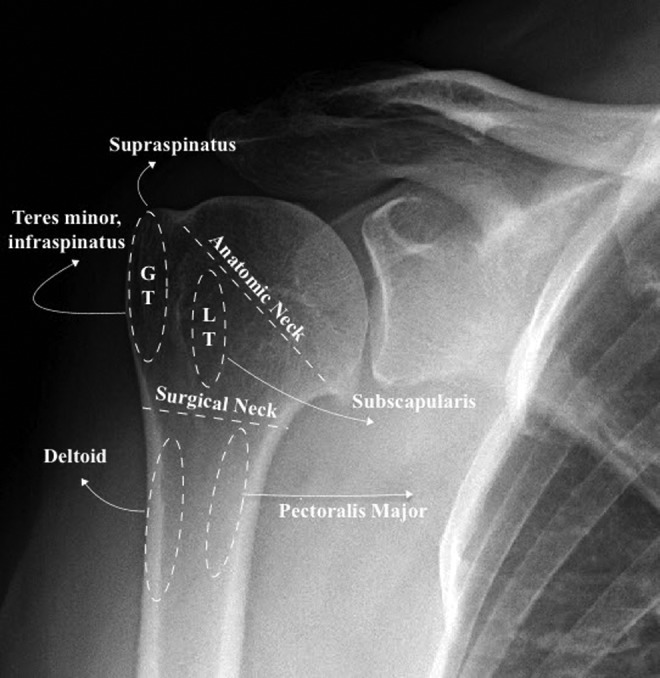
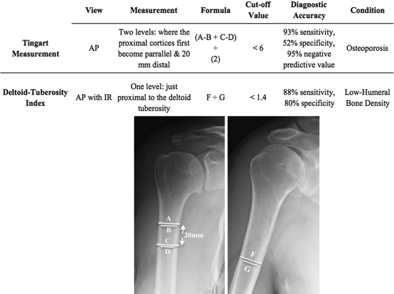
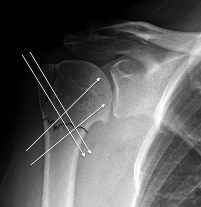
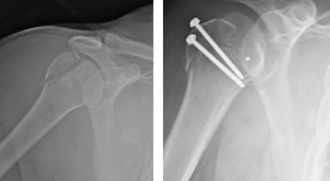
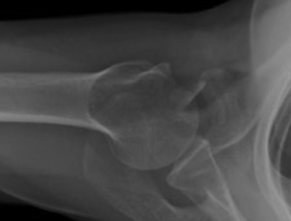
Results: The incidence of proximal humerus fractures is increasing. It is a common osteoporotic fracture. Bone density is a predictor of reduction quality and can be readily assessed with anteroposterior views of the shoulder. Social independence is a predictor of outcome, whereas age is not. Many fractures are minimally displaced and respond acceptably to nonoperative management. Displaced and severe fractures are most frequently treated operatively with intramedullary nails, locking plates, percutaneous techniques, or arthroplasty.
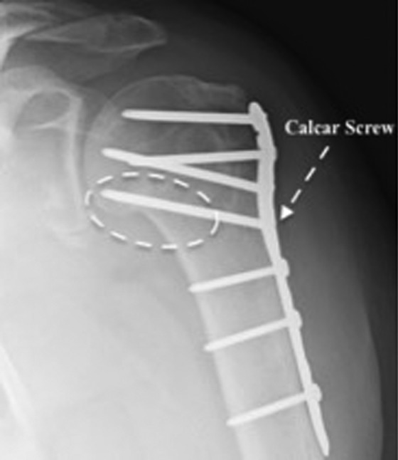
Discussion: Evidence from randomized controlled trials and systematic reviews is insufficient to recommend a treatment; however, most techniques have acceptable or good outcomes. Evaluation should include an assessment of the patient's bone quality, social independence, and surgical risk factors. With internal fixation, special attention should be paid to medial comminution, varus angulation, and restoration of the calcar. With arthroplasty, attention should be paid to anatomic restoration of the tuberosities and proper placement of the prosthesis.
Conclusion: A majority of minimally displaced fractures can be treated conservatively with early physical therapy. Treatment for displaced fractures should consider the patient's level of independence, bone quality, and surgical risk factors. Fixation with percutaneous techniques, intramedullary nails, locking plates, and arthroplasty are all acceptable treatment options. There is no clear evidence-based treatment of choice, and the surgeon should consider their comfort level with various procedures during the decision-making process.
Keywords: fragility fractures; geriatric trauma; osteoporosis; trauma surgery; upper extremity surgery.
Conflict of interest statement
Declaration of Conflicting Interests: The author(s) declared no potential conflicts of interest with respect to the research, authorship, and/or publication of this article.
Figures
References
-
- Court-Brown CM, Caesar B. Epidemiology of adult fractures: a review. Injury. 2006;37(8):691–697. doi:10.1016/j.injury.2006.04.130. - PubMed
-
- Calvo E, Morcillo D, Foruria AM, Redondo-Santamaría E, Osorio-Picorne F, Caeiro JR. Nondisplaced proximal humeral fractures: high incidence among outpatient-treated osteoporotic fractures and severe impact on upper extremity function and patient subjective health perception. J Shoulder Elbow Surg. 2011;20(5):795–801. doi:10.1016/j.jse.2010.09.008. - PubMed
-
- Palvanen M, Kannus P, Niemi S, Parkkari J. Update in the epidemiology of proximal humeral fractures. Clin Orthop. 2006;442:87–92. - PubMed
-
- Savin DD, Zamfirova I, Iannotti J, Goldberg BA, Youderian AR. Survey study suggests that reverse total shoulder arthroplasty is becoming the treatment of choice for four-part fractures of the humeral head in the elderly. Int Orthop. 2016;40(9):1919–1925. doi:10.1007/s00264-016-3227-y. - PubMed
-
- Okike K, Lee OC, Makanji H, Harris MB, Vrahas MS. Factors associated with the decision for operative versus non-operative treatment of displaced proximal humerus fractures in the elderly. Injury. 2013;44(4):448–455. doi:10.1016/j.injury.2012.09.002. - PubMed
Publication types
LinkOut - more resources
Full Text Sources
Other Literature Sources
Medical
