Diagnostic Dilemma: Cerebellopontine Angle Lipoma Versus Dermoid Cyst
- PMID: 29399424
- PMCID: PMC5790211
- DOI: 10.7759/cureus.1894
Diagnostic Dilemma: Cerebellopontine Angle Lipoma Versus Dermoid Cyst
Abstract
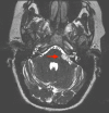
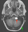
Both lipomas and dermoid cysts of the cerebellopontine angle are rare tumors. These tumors differ in their embryological origin but share similar features on imaging. Both of these congenital lesions can be found in the cerebellopontine angle (CPA), and symptomatic clinical presentation is dictated by the location of the lesion. This paper demonstrates a unique case in which a CPA lipoma was misidentified as a dermoid cyst, leading to surgical intervention. Further, the paper provides a literature review of CPA lipomas and dermoid cysts to aid readers in further differentiating between these two unique tumors.
Keywords: cpa; dermoid cyst; lipoma; posterior fossa; retrosigmoid craniotomy; vertigo.
Conflict of interest statement
The authors have declared that no competing interests exist.
Figures


References
-
- Nonschwannoma tumors of the cerebellopontine angle. Friedmann DR, Grobelny B, Golfinos JG, Roland Jr JT. Otolaryngol Clin North Am. 2015;48:461–475. - PubMed
-
- Imaging of the cerebellopontine angle. Lakshmi M, Glastonbury CM. Neuroimaging Clin N Am. 2009;19:393–406. - PubMed
-
- Cerebellopontine angle masses: radiologic-pathologic correlation. Smirniotopoulos Smirniotopoulos, JG JG, Yue Yue, NC NC, Rushing EJ. Radiographics. 1993;13:1131–1147. - PubMed
-
- Lipomas of the internal auditory canal and cerebellopontine angle. Bacciu A, Di Lella F, Ventura E, Pasanisi E, Russo A, Sanna M. Ann Otol Rhinol Laryngol. 2014;123:58–64. - PubMed
-
- Radiology quiz case 1. Lipoma of the CPA. Klepac N, Hajnsek S, Topic I, Zarkovic K, Ozretic D, Habek M. Arch Otolaryngol Head Neck Surg. 2009;135:828. - PubMed
Publication types
LinkOut - more resources
Full Text Sources
Other Literature Sources
