Review
. 2016 Jul 22:2:119.
doi: 10.21037/jovs.2016.07.10. eCollection 2016.

Endoscopic ultrasound of pancreatic lesions

Affiliations
Review

Endoscopic ultrasound of pancreatic lesions

Charing C N Chong et al. J Vis Surg. .

Abstract

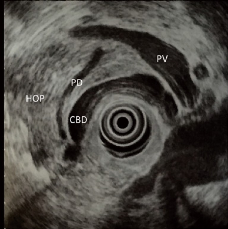
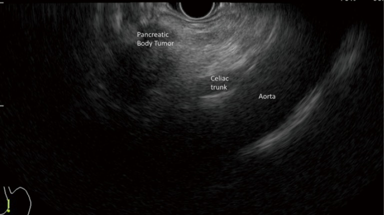
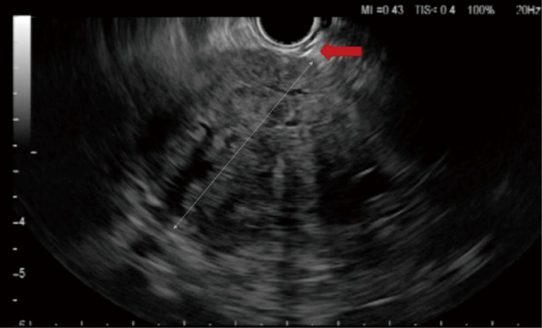
Endoscopic ultrasound (EUS) is a well-established tool for the evaluation of pancreatic lesions. Due to the closer proximity of EUS to the pancreas, EUS offers a high sensitivity for detection of small pancreatic mass and is the preferred modality for obtaining tissue for diagnosis of pancreatic mass. Contrast-enhanced EUS and/or elastography provide additional information to the fundamental B-mode ultrasound images, leading to more accurate diagnosis. The aim of this video-article is to show the different steps in performing EUS on pancreatic lesions and to provide some tips and tricks to improve and facilitate the execution of EUS on pancreatic lesions.

Keywords: Endoscopic ultrasound (EUS); endoscopic ultrasonography; fine needle aspiration (FNA); pancreas.


Conflict of interest statement

Conflicts of Interest: The authors have no conflicts of interest to declare.

Figures

Figure 1
Figure 1
Radial endoscopic ultrasound (EUS) image of pancreatic head. PV, portal vein; CBD, common bile duct; PD, pancreatic duct; HOP, head of pancreas.
Figure 2
Figure 2
Image of linear endoscopic ultrasound (EUS) showing a pancreatic body tumor and its relationship with aorta and celiac trunk.
Figure 3
Figure 3
Examination of pancreas with radial endoscopic ultrasound (EUS) (6). Available online: http://www.asvide.com/articles/1042
Figure 4
Figure 4
Examination of pancreas with linear endoscopic ultrasound (EUS) (7). Available online: http://www.asvide.com/articles/1043
Figure 5
Figure 5
Video showing endoscopic ultrasound (EUS) fine needle aspiration (FNA) of a pancreatic body tumor (8). Available online: http://www.asvide.com/articles/1044
Figure 6
Figure 6
Image of linear endoscopic ultrasound (EUS) showing the sheath of fine needle aspiration (FNA) needle protruding out from the channel, indicated by the red arrow.
Figure 7
Figure 7
Video illustration on the detection and characterization of a small pancreatic cancer by a combination of conventional endoscopic ultrasound (EUS), contrast enhanced harmonic EUS (CEH-EUS) and elastography (15). Available online: http://www.asvide.com/articles/1045

References

    1. DiMagno EP, Buxton JL, Regan PT, et al. Ultrasonic endoscope. Lancet 1980;1:629-31. 10.1016/S0140-6736(80)91122-8 - DOI - PubMed
    1. Yasuda K, Tanaka Y, Fujimoto S, et al. Use of endoscopic ultrasonography in small pancreatic cancer. Scand J Gastroenterol Suppl 1984;102:9-17. - PubMed
    1. DeWitt J, Devereaux B, Chriswell M, et al. Comparison of endoscopic ultrasonography and multidetector computed tomography for detecting and staging pancreatic cancer. Ann Intern Med 2004;141:753-63. 10.7326/0003-4819-141-10-200411160-00006 - DOI - PubMed
    1. Gress FG, Hawes RH, Savides TJ, et al. Role of EUS in the preoperative staging of pancreatic cancer: a large single-center experience. Gastrointest Endosc 1999;50:786-91. 10.1016/S0016-5107(99)70159-8 - DOI - PubMed
    1. Shrikhande SV, Barreto SG, Goel M, et al. Multimodality imaging of pancreatic ductal adenocarcinoma: a review of the literature. HPB (Oxford) 2012;14:658-68. 10.1111/j.1477-2574.2012.00508.x - DOI - PMC - PubMed

LinkOut - more resources