Review
. 2017 Dec;7(Suppl 3):S159-S164.
doi: 10.21037/cdt.2017.10.14.

May-Thurner: diagnosis and endovascular management

Affiliations
Review

May-Thurner: diagnosis and endovascular management

M-Grace Knuttinen et al. Cardiovasc Diagn Ther. 2017 Dec.

Abstract

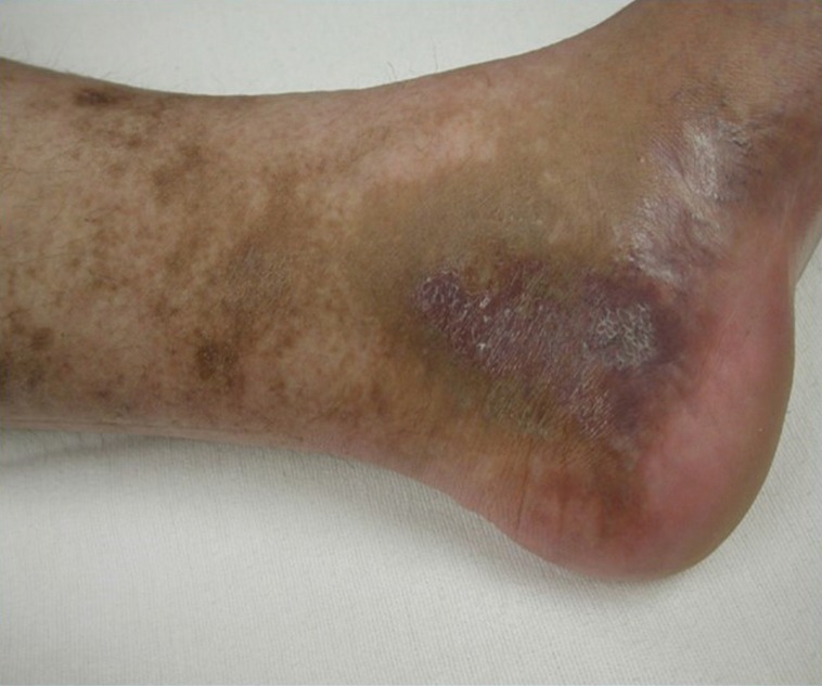
Common left iliac vein compression, otherwise known as May-Thurner (MT), is an anatomical risk factor for lower extremity deep vein thrombosis (DVT). MT refers to chronic compression of the left iliac vein against the lumbar spine by the overlying right common iliac artery. The compression may be asymptomatic. The syndrome is a clinical spectrum of physical findings and history plus the lesion. It is characterized by the varying degrees of venous hypertension. This can be non-thrombotic, combined with acute DVT or post-thrombotic. Traditionally, acute DVT was treated with standard anticoagulation and sometimes, thrombectomy. However these measures do not address the underlying culprit lesion of mechanical compression. Furthermore, if managed only with anticoagulation, patients with residual thrombus are at risk for developing recurrent DVT or post-thrombotic syndrome (PTS). Both retrospective and prospective studies have shown that endovascular management should be the preferred approach to dissolve proximal thrombus and to also treat the underlying compression with endovascular stent placement.

Keywords: May-Thurner (MT); deep vein thrombosis (DVT); stent.


Conflict of interest statement

Conflicts of Interest: The authors have no conflicts of interest to declare.

Figures

Figure 1
Figure 1
Clinical example of patient presenting with left lower extremity swelling related to MTS. MTS, May-Thurner syndrome.
Figure 2
Figure 2
The clinical example of venous ulceration and hyperpigmentation that can occur in patients with MTS. MTS, May-Thurner syndrome.
Figure 3
Figure 3
Illustration of May-Thurner as seen on venography and intravascular ultrasound. (A) Left lower extremity venogram shows the compression of the right common iliac artery (arrow) as it courses across the left common iliac vein. Collaterals are also noted; (B) correspondent intravascular images show the markedly narrowed compressed left iliac vein.
Figure 4
Figure 4
The post stent venogram shows complete patency of the left common iliac vein with good forward flow into the IVC. Correspondent intravascular ultrasound (IVUS) shows the resultant normalized caliber of the left common iliac vein following stent placement. Excellent stent apposition is noted on IVUS. IVC, inferior vena cava.

References

    1. May R, Thurner J. The cause of the predominantly sinistral occurrence of thrombosis of the pelvic veins. Angiology 1957;8:419-27. 10.1177/000331975700800505 - DOI - PubMed
    1. Oğuzkurt L, Ozkan U, Tercan F, et al. Ultrasonographic diagnosis of iliac vein compression (May-Thurner) syndrome. Diagn Interv Radiol 2007;13:152-5. - PubMed
    1. Kibbe MR, Ujiki M, Goodwin AL, et al. Iliac vein compression in an asymptomatic patient population. J Vasc Surg 2004;39:937-43. 10.1016/j.jvs.2003.12.032 - DOI - PubMed
    1. Raju S, Neglen P. High prevalence of nonthrombotic iliac vein lesions in chronic venous disease: a permissive role in pathogenicity. J Vasc Surg 2006;44:136-43;discussion 144. 10.1016/j.jvs.2006.02.065 - DOI - PubMed
    1. Kasirajan K, Gray B, Ouriel K. Percutaneous AngioJet thrombectomy in the management of extensive deep venous thrombosis. J Vasc Interv Radiol 2001;12:179-85. 10.1016/S1051-0443(07)61823-5 - DOI - PubMed

LinkOut - more resources