. 2018 Sep;12(3):575-588.
doi: 10.1007/s12079-018-0456-4. Epub 2018 Feb 4.

Oxygen drives hepatocyte differentiation and phenotype stability in liver cell lines

Affiliations

Oxygen drives hepatocyte differentiation and phenotype stability in liver cell lines

Martien van Wenum et al. J Cell Commun Signal. 2018 Sep.

Abstract

The in vitro generation of terminally differentiated hepatocytes is an unmet need. We investigated the contribution of oxygen concentration to differentiation in human liver cell lines HepaRG and C3A. HepaRG cells were cultured under hypoxia (5%O2), normoxia (21%O2) or hyperoxia (40%O2). Cultures were analysed for hepatic functions, gene transcript levels, and protein expression of albumin, hepatic transcription factor CEBPα, hepatic progenitor marker SOX9, and hypoxia inducible factor (HIF)1α. C3A cells were analysed after exposure to normoxia or hyperoxia. In hyperoxic HepaRG cultures, urea cycle activity, bile acid synthesis, CytochromeP450 3A4 (CYP3A4) activity and ammonia elimination were 165-266% increased. These effects were reproduced in C3A cells. Whole transcriptome analysis of HepaRG cells revealed that 240 (of 23.223) probes were differentially expressed under hyperoxia, with an overrepresentation of genes involved in hepatic differentiation, metabolism and extracellular signalling. Under hypoxia, CYP3A4 activity and ammonia elimination were inhibited almost completely and 5/5 tested hepatic genes and 2/3 tested hepatic transcription factor genes were downregulated. Protein expression of SOX9 and HIF1α was strongly positive in hypoxic cultures, variable in normoxic cultures and predominantly negative in hyperoxic cultures. Conversely, albumin and CEBPα expression were highest in hyperoxic cultures. HepaRG cells that were serially passaged under hypoxia maintained their capacity to differentiate under normoxia, in contrast to cells passaged under normoxia. Hyperoxia increases hepatocyte differentiation in HepaRG and C3A cells. In contrast, hypoxia maintains stem cell characteristics and inhibits hepatic differentiation of HepaRG cells, possibly through the activity of HIF1α.

Keywords: C3A; HepaRG; Hepatic progenitor cell; Hepatocyte differentiation; Oxygen; Propagation capacity.


Conflict of interest statement

RC and RH are inventors of the patent-protected HepaRG-based AMC-Bioartificial liver that is currently being commercialized through the university spin-off company Hep-Art Medical Devices B.V of which RC is unsalaried Chief Scientific Officer. RH was previously employed part-time by Hep-Art Medical Devices B.V.

The authors have no other relevant affiliations or financial involvement with any organization or entity with a financial interest in or financial conflict with the subject matter or materials discussed in the manuscript apart from those disclosed.

Figures

Fig. 1
Fig. 1
Ambient oxygen concentration influences HepaRG cell morphology but not growth. a Pericellular oxygen concentration in culture medium with- and without 4-week old HepaRG cultures and (b) total protein content of 4-week old cultures. There was a marked difference in morphology of HepaRG cells cultured under: (c) normoxia, (d) hypoxia or (e) hyperoxia. Scale bar = 100 μM
Fig. 2
Fig. 2
Nuclear SOX9 expression is downregulated, while CEBPα is upregulated at increased oxygen levels. a HepaRG cells cultured under normoxia (top), hypoxia (middle) and hyperoxia (bottom), stained for: DAPI (blue), Albumin (green) and SOX9 (red). Arrows A&B indicate nuclear translocation of SOX9 in HepaRG-Hypoxia, and to a lesser degree in HepaRG-Normoxia. Arrow C indicates mainly cytosolic SOX9 expression in HepaRG-Hyperoxia, with SOX9-negative nuclei. b HepaRG cells cultured under normoxia (top), hypoxia (middle) and hyperoxia (bottom), stained for: DAPI (blue) and CEBPα (red). Arrow A indicates positive nuclear staining. Scale bar = 50 μM
Fig. 2
Fig. 2
Nuclear SOX9 expression is downregulated, while CEBPα is upregulated at increased oxygen levels. a HepaRG cells cultured under normoxia (top), hypoxia (middle) and hyperoxia (bottom), stained for: DAPI (blue), Albumin (green) and SOX9 (red). Arrows A&B indicate nuclear translocation of SOX9 in HepaRG-Hypoxia, and to a lesser degree in HepaRG-Normoxia. Arrow C indicates mainly cytosolic SOX9 expression in HepaRG-Hyperoxia, with SOX9-negative nuclei. b HepaRG cells cultured under normoxia (top), hypoxia (middle) and hyperoxia (bottom), stained for: DAPI (blue) and CEBPα (red). Arrow A indicates positive nuclear staining. Scale bar = 50 μM
Fig. 3
Fig. 3
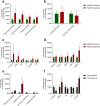
Ambient hypoxia augments hepatic differentiation of HepaRG and C3A cells. HepaRG monolayers cultured under ambient normoxia or hyperoxia were tested for hepatic functions (a), glucose consumption and lactate production (b), as well as transcript levels of hepatic genes (c) and transcription factor genes (d). C3A monolayers cultured under normoxia and hyperoxia were tested for hepatic functions (e) and hepatic gene transcript levels (f). * = P < 0.05 compared to HepaRG normoxia
Fig. 4
Fig. 4
Whole-transcriptome microarray analysis on HepaRG cells cultured under normoxia or hyperoxia and primary human hepatocytes. Venn diagram of differentially expressed genes (adj. P < 0.05) between freshly isolated PHHs and HepaRG cells cultured under ambient normoxia or hyperoxia
Fig. 5
Fig. 5
HepaRG cells cultured under hypoxia lose hepatic gene transcription and –functionality. HepaRG monolayers cultured under ambient normoxia or hypoxia were tested for hepatic functions (a), glucose consumption and lactate production (b), as well as transcript levels of hepatic genes (c) and transcription factor genes (d). * = P < 0.05 compared to HepaRG normoxia
Fig. 6
Fig. 6
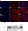
Nuclear HIF1α expression decreases at increased oxygen levels. a HepaRG cells cultured under normoxia (top), hypoxia (middle) and hyperoxia (bottom), stained for: DAPI (blue) and HIF1α (red). Arrow A indicates nuclear expression of HIF1α, arrow B indicates cells that appear free of nuclear HIF1α. Scale bar = 50 μM. b Western blot showing expression of HIF1α (top lanes), and vinculin as loading control (bottom lanes). Targets and positive controls are parts of the same image
Fig. 7
Fig. 7
Expansion under hypoxia stabilizes the HepaRG phenotype during serial passaging. HepaRG cells at passage 17 (P17) from isolation were split into sub lines that were maintained under ambient hypoxia or normoxia. Cultures were passaged every other week, every other passage samples from both cultures were cultured under normoxia for 4 weeks and tested (a). Cultures were tested on ammonia elimination (b), total protein content (c), transcript levels of hepatic genes (d) and transcription factor genes (e), lactate production (f) and glucose consumption (g). * = P < 0.05 compared to normoxia maintained line of same passage number. $ = P < 0.05 compared to P17

References

    1. Amarilio R, Viukov SV, Sharir A, et al. HIF1alpha regulation of Sox9 is necessary to maintain differentiation of hypoxic prechondrogenic cells during early skeletogenesis. Development. 2007;134(21):3917–3928. doi: 10.1242/dev.008441. - DOI - PubMed
    1. Ashburner M, Ball CA, Blake JA, et al. Gene ontology: tool for the unification of biology. The Gene Ontology Consortium. Nat Genet. 2000;25(1):25–29. doi: 10.1038/75556. - DOI - PMC - PubMed
    1. Boulter L, Govaere O, Bird TG, et al. Macrophage-derived Wnt opposes Notch signaling to specify hepatic progenitor cell fate in chronic liver disease. Nat Med. 2012;18(4):572–579. doi: 10.1038/nm.2667. - DOI - PMC - PubMed
    1. Buck LD, Inman SW, Rusyn I, et al. Co-regulation of primary mouse hepatocyte viability and function by oxygen and matrix. Biotechnol Bioeng. 2014;111(5):1018–1027. doi: 10.1002/bit.25152. - DOI - PMC - PubMed
    1. Cerec V, Glaise D, Garnier D, et al. Transdifferentiation of hepatocyte-like cells from the human hepatoma HepaRG cell line through bipotent progenitor. Hepatology. 2007;45(4):957–967. doi: 10.1002/hep.21536. - DOI - PubMed