. 2018;8(2):303-322.
doi: 10.3233/JPD-171248.

Best Practices for Generating and Using Alpha-Synuclein Pre-Formed Fibrils to Model Parkinson's Disease in Rodents

Affiliations

Best Practices for Generating and Using Alpha-Synuclein Pre-Formed Fibrils to Model Parkinson's Disease in Rodents

Nicole K Polinski et al. J Parkinsons Dis. 2018.

Abstract

Parkinson's disease (PD) is the second most common neurodegenerative disease, affecting approximately one-percent of the population over the age of sixty. Although many animal models have been developed to study this disease, each model presents its own advantages and caveats. A unique model has arisen to study the role of alpha-synuclein (aSyn) in the pathogenesis of PD. This model involves the conversion of recombinant monomeric aSyn protein to a fibrillar form-the aSyn pre-formed fibril (aSyn PFF)-which is then injected into the brain or introduced to the media in culture. Although many groups have successfully adopted and replicated the aSyn PFF model, issues with generating consistent pathology have been reported by investigators. To improve the replicability of this model and diminish these issues, The Michael J. Fox Foundation for Parkinson's Research (MJFF) has enlisted the help of field leaders who performed key experiments to establish the aSyn PFF model to provide the research community with guidelines and practical tips for improving the robustness and success of this model. Specifically, we identify key pitfalls and suggestions for avoiding these mistakes as they relate to generating the aSyn PFFs from monomeric protein, validating the formation of pathogenic aSyn PFFs, and using the aSyn PFFs in vivo or in vitro to model PD. With this additional information, adoption and use of the aSyn PFF model should present fewer challenges, resulting in a robust and widely available model of PD.

Keywords: Alpha-synuclein; Parkinson’s disease; pre-formed fibril; preclinical model.


Figures

Fig.1
Fig.1
Schematic depiction of the protocol for generating alpha-synuclein pre-formed fibrils (aSyn PFFs) from specially formulated monomers. The protocol for generating aSyn PFFs from monomers includes three main stages, (A) preparation of aSyn monomers, (B) generation of aSyn PFFs from monomers, and (C) preparation of aSyn PFFS for use. A) The protocol begins with the monomeric recombinant aSyn protein specially formulated for aSyn PFF development, which is then centrifuged for the supernatant to be transferred and protein concentration to be measured. B) To generate aSyn PFFs, the solution is diluted, vortexed, and incubated at 37°C shaking for 7 days before quality control and storage. Please note that a portion of the monomeric starting material should be set aside at the beginning of this step for use as a negative control in quality control experiments, with even larger quantities set aside if monomers are to be used as the control protein in model generation. C) On the day of use, an aliquot of aSyn PFFs should be thawed, diluted after the protein concentration has been re-measured, sonicated, and quality controlled prior to use. The protocol corresponding to this schematic can be found in the Supplementary Material. aSyn, alpha-synuclein; PFF, pre-formed fibril; s, seconds; d, days; RT, room temperature.
Fig.2
Fig.2
Impact of storage conditions on alpha-synuclein pre-formed fibril (aSyn PFF) structure and pathogenicity. A-C) Representative transmission electron microscopy (TEM) images of aSyn PFF samples stored at varying temperatures before sonication. A) Samples stored at room temperature for 3–4 weeks display long fibrillar structures characteristic of unsonicated aSyn PFFs. B) Samples stored at –80°C for 4–5 months display long fibril structures as well as fractured, smaller aggregates. C) Samples stored in liquid nitrogen for 4–5 months display a distinct morphology characterized by fractured fibrils and non-specific aggregates (arrowhead). D–F) Analysis of the impact of storage conditions on aSyn PFF pathogenicity in primary hippocampal neuron cultures. Primary hippocampal neurons were exposed to fibrils at 0.2 μg/mL and fixed 6 days later. D–E) Immunofluorescence was performed using an antibody to pS129 aSyn to visualize inclusions (green) or to neurofilament heavy chain (blue) to visualize axons. Scale bars = 50 μm. Fibrils were either (D) generated immediately before adding to primary neurons or (E) stored at –80°C for 6 months before use. F) Quantitation of the percent area occupied by pS129 aSyn immunoreactivity reveals a highly significant (p < 0.001) difference between fresh and frozen aSyn PFFs, with much greater levels of pS129 aSyn induced by fresh aSyn PFFs as compared to frozen aSyn PFFs. Graph depicts mean values with error bars denoting standard deviation. aSyn, alpha-synuclein; PFF, pre-formed fibril; RT, room temperature; liquid N2, liquid nitrogen; p-α-syn, pS129 aSyn.
Fig.3
Fig.3
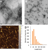
Analysis of the size and morphology of alpha-synuclein pre-formed fibrils (aSyn PFFs) after efficient sonication. A-B) Transmission electron microscopy (TEM) images of aSyn PFFs (A) pre-sonication and (B) post-sonication. A) aSyn PFFs pre-sonication are long fibrils with beta-sheet structure. Scale bars = 50 nm. B) aSyn PFFs that have been efficiently sonicated are short fibrils that keep the beta-sheet conformation. Scale bars = 50 nm. C) Atomic force microscopy (AFM) analysis of sonicated aSyn PFFs to determine size of the sonicated PFFs. D) Size distribution of the aSyn PFFs after sonication as determined by values obtained from statistical analysis of the aggregates identified in the atomic force microscopy images (C). aSyn, alpha-synuclein; PFF, pre-formed fibril.
Fig.4
Fig.4
Schematic depiction of the workflow for generating, validating, and using alpha-synuclein pre-formed fibrils (aSyn PFFs) as the experimental protein and aSyn monomers as the control protein. When using aSyn PFFs as the experimental protein and aSyn monomers as the control, the first step for preparation and use of both samples is to defrost the specially-formulated aSyn monomers for PFF generation on ice and to preform endotoxin cleanup, if required (Green box). Once thawed, the sample should be divided in two, with half designated for use as the monomeric control protein (Blue boxes) and the other half destined for generation of PFFs (Orange boxes). Protocols for generating aSyn PFFs are located in the Supplementary Material, Fig. 1, and the cited literature. After the PFFs have been generated, aliquot into single use tubes of 5 mg/ml and store at –80°C for long-term storage or room temperature for short term storage. Use one aliquot to validate proper fibril formation (see Fig. 5A–C). Once fibril formation has been confirmed, sonication parameters must be validated. For this, use one aliquot (if stored at –80°C, defrost and keep at room temperature), dilute to the desired working concentration, and sonicate. After sonication, the fibril size and pathogenicity must be validated (see Fig. 5D–G). Similarly, aSyn monomers for the control samples should be aliquotted into single-use tubes at ≤7.5 mg/ml, validated alongside the aSyn PFFs as the control, and stored at –80°C. These validation steps should be performed in advance of use in order to confirm proper sample composition and to verify sonication parameters. On the day of use, defrost one aliquot of the aSyn PFFS (thaw at room temperature if frozen) and aSyn monomers (thaw on ice). Prepare using the validated dilutions and—in the case of the aSyn PFFs—sonication parameters tested previously. At this step it may be wise to again confirm sample composition via electron microscopy before use. Keep the aSyn PFFs at room temperature and the aSyn monomers on ice during use. Mix solutions between injections to ensure the larger aggregates do not pellet and lead to sample heterogeneity. A sample can be used for four hours before it should be replaced by a new sample. aSyn, alpha-synuclein; PFF, pre-formed fibrils; RT, room temperature; EM, electron microscopy; hrs, hours.
Fig.5
Fig.5
Recommended quality control experiments to verify proper alpha-synuclein (aSyn) fibril formation and preparation. Depiction of recommended quality control experiments (denoted with *) or supplemental quality control experiments with expected results. A–C) After the aSyn PFFs are generated from the monomeric starting material (Fig. 1B), samples should visually appear opaque and should be validated to confirm (A) amyloid conformation of the fibrils and (B–C) formation of long fibrillar protein aggregates. These validation efforts should be performed with every new batch of aSyn PFFs. A) The thioflavin T (ThT) assay is recommended to confirm presence of beta-sheet structures. Expected results include high levels of ThT fluorescent signal with PFFs as compared to monomers. It should be noted that human aSyn PFFs will elicit a stronger ThT signal than mouse aSyn PFFs [51]. B) The sedimentation assay is recommended to confirm aggregate formation in aSyn PFF samples. Expected results include substantially more protein in the pellet (pel) fraction as compared to the supernatant (sup) fraction for aSyn PFFs and the opposite result for monomeric aSyn protein. C) If sedimentation assays are not feasible or additional quality control is desired, electron microscopy is recommended to visualize the aSyn PFFs. Electron microscopy results should primarily show elongated fibrils. Scale bars = 50 nm. D–H) aSyn PFFs should again be validated post-sonication (Fig. 1C) to confirm (D–E) seeding capacity, (F–G) proper size of sonicated aSyn PFFs, and (H) in vivo pathogenicty. These validation efforts are at a minimum recommended when establishing or changing sonication parameters or before long-term in vivo studies. D–E) Seeding capacity should be confirmed using either (D) in vitro seeding experiments or (E) the ThT kinetic assay. D) In vitro seeding experiments are recommended for confirming the pathogenicity of aSyn fibrils as this will model the conversion of endogenous aSyn into pS129 aSyn in neurons after incubation with aSyn PFFs. Expected results include high levels of pS129 staining post-incubation with aSyn PFFs but no appreciable pS129 aSyn staining post-incubation with monomers or inadequately sonicated PFFs. Scale bar = 50 μm. E) If in vitro seeding assays are not feasible, the ThT kinetic assay may be used to confirm seeding capacity of aSyn PFF samples. Expected results include no increase in ThT fluorescence with addition of monomers and an increasing rate of ThT fluorescence with increased sonication time, indicating more pathogenic fibrils [35]. This assay is recommended when comparing different sonication parameters or when used with a positive control as the rate of increase or peak fluorescence levels may vary between runs and comparing only sonicated PFFs to monomers will not be informative. F–G) Average fibril size should also be confirmed post-sonication and before use by either (F) electron microscopy or (G) dynamic light scattering (DLS). F) Electron microscopy is recommended for visualizing sonicated aSyn PFFs to confirm size and uniformity of aggregates. The majority of fibrils analyzed should be 50 nm or smaller to elicit high levels of pathology (graph inset indicates average fibril size). Scale bars = 50 nm. G) If electron microscopy is not feasible or additional quality control is desired, DLS may be used to analyze fibril size [35]. Again, the majority of fibrils should be 50 nm or smaller to properly seed pathology. This graph depicts DLS data for fibrils separated by size. H) If using aSyn PFFs in a long-term in vivo study, it is highly recommended to perform a short term in vivo pilot study to verify in vivo pathogenicity and injection parameters. Thirty days post-injection may be sufficient to visualize early aSyn pathology by pS129 aSyn staining in the mouse [51] or rat [34] brain following striatal injection of mouse aSyn PFFs, although longer time points may be required. sup, supernatant; pel, pellet; aSyn, alpha-synuclein; PFF, pre-formed fibril; ThT, thioflavin T; AU, arbitrary units; sec, seconds; p-α-syn, alpha-synuclein phosphorylated at S129; M.W., molecular weight.
Fig.6
Fig.6
Optimized striatal injection coordinates for use in the rat alpha-synuclein pre-formed fibril (aSyn PFF) model. A–B) Comparison of pS129 aSyn expression in the ipsilateral midbrain after two site striatal injections using (A) original, traditionally-used coordinates [34] or (B) optimized coordinates guided by recent findings revealing distinct SNpc-striatal innervation patterns [54]. Neuroanatomical atlas images from Paxinos and Watson [58] depict the general coordinates used for injection. Representative low and high magnification images are shown for ipsilateral midbrain stained with cresyl violet (purple) and antibodies directed against pS129 aSyn (brown). A) Traditional injection coordinates involve injection into the dorsomedial striatum (DMS) and ventrolateral striatum (VLS). Injection into these coordinates induces robust pS129 aSyn expression in the SNpc as well as the ventral tegmental area (VTA). B) Optimized injection coordinates involve injection into the DMS as well as the dorsolateral striatum (DLS). Injection into these coordinates induces robust pS129 aSyn expression that is localized primarily to the SNpc and absent from the VTA. C–D) Quantitation of the percentage of pS129 aggregates in the (C) SNpc or (D) VTA following injection using the original (DMS + VLS) or optimized (DMS + DLS) injection coordinates. Injection using the original coordinates results in ∼80% of aggregates in the SNpc and ∼20% of aggregates in the VTA whereas injection using the optimized coordinates results in ∼90% of aggregates in the SNpc and ∼10% of aggregates in the VTA. Mean values are shown with error bars denoting standard error of the mean. aSyn, alpha-synuclein; PFF, pre-formed fibril; DMS, dorsomedial striatum; VLS, ventrolateral striatum; DLS, dorsolateral striatum; SNpc, substantia nigra pars compacta; VTA, ventral tegemental area; pS129 aSyn, alpha-synuclein phosphorylated at S129.
Fig.7
Fig.7
Common pitfalls in the alpha-synuclein pre-formed fibril (aSyn PFF) model and solutions for avoiding these mistakes. Six common issues that lead to lack of pathology or other issues in the aSyn PFF model include (A) using an incompatible protein as the monomeric starting material or injected material, (B) forming PFFs in an incompatible buffer, (C) storing or keeping solutions at the incorrect temperature, (D) inadequately sonicating the aSyn PFF sample, (E) failing to validate the aSyn PFFs have the proper biophysical and biochemical properties, and (F) choosing an unideal control or not accounting for the control when reserving aSyn monomeric protein. These are six very important factors to which attention should be paid and caution should be taken to avoid. Guidelines for how to avoid these pitfalls are included as well. aSyn, alpha-synuclein; PFF, pre-formed fibril; RT, room temperature.

References

    1. Dorsey ER, Constantinescu R, Thompson JP, Biglan KM, Holloway RG, Kieburtz K, Marshall FJ, Ravina BM, Schifitto G, Siderowf A, Tanner CM (2007) Projected number of people with Parkinson disease in the most populous nations, 2005 through 2030. Neurology 68, 384–386. - PubMed
    1. Graybiel AM, Hirsch EC, Agid Y (1990) The nigrostriatal system in Parkinson’s disease. Adv Neurol 53, 17–29. - PubMed
    1. Burke RE, O’Malley K (2013) Axon degeneration in Parkinson’s disease. Exp Neurol 246, 72–83. - PMC - PubMed
    1. Pollanen MS, Dickson DW, Bergeron C (1993) Pathology and biology of the Lewy body. J Neuropathol Exp Neurol 52, 183–191. - PubMed
    1. Spillantini MG, Schmidt ML, Lee VM, Trojanowski JQ, Jakes R, Goedert M (1997) Alpha-synuclein in Lewy bodies. Nature 388, 839–840. - PubMed

Substances