. 2018 Mar;24(3):368-374.
doi: 10.1038/nm.4485. Epub 2018 Feb 5.

Congenital Zika virus infection as a silent pathology with loss of neurogenic output in the fetal brain

Affiliations

Congenital Zika virus infection as a silent pathology with loss of neurogenic output in the fetal brain

Kristina M Adams Waldorf et al. Nat Med. 2018 Mar.

Abstract

Zika virus (ZIKV) is a flavivirus with teratogenic effects on fetal brain, but the spectrum of ZIKV-induced brain injury is unknown, particularly when ultrasound imaging is normal. In a pregnant pigtail macaque (Macaca nemestrina) model of ZIKV infection, we demonstrate that ZIKV-induced injury to fetal brain is substantial, even in the absence of microcephaly, and may be challenging to detect in a clinical setting. A common and subtle injury pattern was identified, including (i) periventricular T2-hyperintense foci and loss of fetal noncortical brain volume, (ii) injury to the ependymal epithelium with underlying gliosis and (iii) loss of late fetal neuronal progenitor cells in the subventricular zone (temporal cortex) and subgranular zone (dentate gyrus, hippocampus) with dysmorphic granule neuron patterning. Attenuation of fetal neurogenic output demonstrates potentially considerable teratogenic effects of congenital ZIKV infection even without microcephaly. Our findings suggest that all children exposed to ZIKV in utero should receive long-term monitoring for neurocognitive deficits, regardless of head size at birth.


Conflict of interest statement

Competing Financial Interests

The authors declare no competing financial interests.

Figures
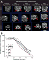
Figure 1
Figure 1
Fetal Brain MRI Imaging and Volume Analysis. Serial fetal brain MRI images (HASTE) from pigtail macaques inoculated with ZIKV and control media were analyzed for differences in structure and volume. Four of five ZIKV animals demonstrated periventricular-subcortical T2-hyperintense foci in the posterior brain between 120–129 days, which were absent in control fetuses at the same developmental age (A). Images were segmented to obtain specific brain volumes of each region (e.g. white matter, cortical gray matter, Fig. S13). The plot (B) demonstrates the change in the supratentorial (fetal brain) tissue volume ratio in the latter half of pregnancy; this ratio estimates the contribution of non-cortical tissues (excluding cortical plate) to the overall volume of the brain (excluding cerebellum).
Figure 2
Figure 2
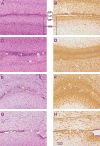
Neuropathology of the Fetal Brain Demonstrating ZIKV-Associated Ependymal Fusion and Periventricular Gliosis. Microscopic images of hematoxylin and eosin stained sections of the lateral ventricle (LV) with ependymal epithelium (EP) are shown for a control (A) and ZIKA 1, 2 and 4 (C, E, G, respectively). Immunostaining for GFAP is also shown in the same control (B) and ZIKV-infected animals (D, F, H) in an adjacent section. Asterisks indicate zones of ependymal fusion. GFAP-immunostaining demonstrates periventricular gliosis (brown staining) in regions surrounding ependymal fusion. WM, white matter.
Figure 3
Figure 3
Reduced NSC proliferation in late fetal neurogenic zones. A schematic of the fetal temporal cortical subventricular zone (SVZ) and hippocampus is shown for a rhesus macaque neonate (0 months) with a Nissl stained section (A) and diagrams to indicate locations of proliferating Ki67+ NSCs (pink dots and pink regions) in neurogenic niches of the tissue (B) and specific regions of the SVZ and hippocampus (C). Ki67+ cells in the dentate gyrus and the SVZ, key regions of neurogenesis, are shown for a control (row D) and ZIKA fetuses (E–G), with higher power views of the dentate gyrus (blue box) and SVZ regions (pink box) to the right. (H) Quantitation of Ki67+ cells reveals a significant decrease in proliferation in the SVZ of ZIKV-infected animals compared to SGZ and controls. The dentate gyrus zones include the molecular layer (ML), granule zone (GZ), subgranular zone (SGZ), polymorphic layer (PM), and the hilus (H). Adjacent areas to the dentate include the cornu ammonis (CA) and subiculum. The lateral ventricle (LV) is lined by ependymal epithelium (E) with an underlying SVZ. Data shown as mean ± standard deviation of Ki67+ cell counts per mm2 (*p<0.05). The schematic (A) is based on the NIH Blueprint Nonhuman Primate Atlas (http://www.blueprintnhpatlas.org).
Figure 4
Figure 4
Attenuated neurogenesis in late fetal neurogenic zones accompanied by granule neuron dysmorphia in the dentate gyrus after maternal ZIKV infection. (A–C) Adjacent sections were labeled for Sox2+ NSCs (left sections) and Tbr2+ IPs (right sections) by immunohistochemistry (brown, Tbr2 is also known as Eomes) and quantified by image analysis (D, E). Fetal Sox2+ NSCs were significantly reduced in the SVZ and disorganized in the subgranular zone (SGZ) with congenital ZIKV exposure. In the SGZ, there was a significant and marked reduction in Tbr2+ IPs in ZIKA fetuses compared to controls. Asterisks indicate disordered NSC in the SGZ neurogenic niche. Arrows indicate a loss of continuity in the granule zone. (F–H) Confocal microscopy to identify NSCs (Sox2+, red), IPs (Tbr2+, green) and immature granule neurons (Dcx, doublecortin, white; Dapi nuclei, blue) in adjacent sections revealed disordered NSCs in the SGZ niche, loss of neurogenic output (Tbr2+ IPs), and dysmorphic granule neurons with congenital ZIKV exposure. Data shown as mean ± standard deviation of either Sox2+ or Tbr2+ cell counts per mm2 (**p<0.01).

Comment in

References

    1. van den Pol AN, Mao G, Yang Y, Ornaghi S, Davis JN. Zika virus targeting in the developing brain. J Neurosci. 2017 - PMC - PubMed
    1. Moore CA, et al. Characterizing the Pattern of Anomalies in Congenital Zika Syndrome for Pediatric Clinicians. JAMA Pediatr. 2016 - PMC - PubMed
    1. Maurice J. The Zika virus public health emergency: 6 months on. Lancet. 2016;388:449–450. - PubMed
    1. Franca GVA, et al. Congenital Zika virus syndrome in Brazil: a case series of the first 1501 livebirths with complete investigation. Lancet. 2016;388:891–897. - PubMed
    1. Mlakar J, et al. Zika Virus Associated with Microcephaly. N Engl J Med. 2016 - PubMed

Publication types

MeSH terms