. 2018 Sep 8;218(8):1272-1283.
doi: 10.1093/infdis/jiy062.

Vaccine-Associated Maintenance of Epithelial Integrity Correlated With Protection Against Virus Entry

Affiliations

Vaccine-Associated Maintenance of Epithelial Integrity Correlated With Protection Against Virus Entry

L Shang et al. J Infect Dis. .

Abstract

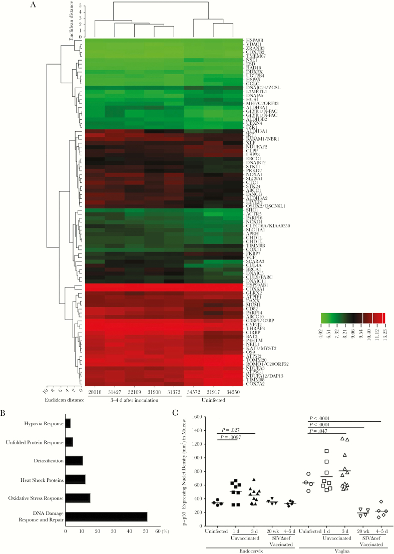
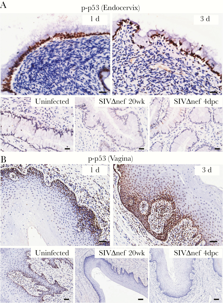
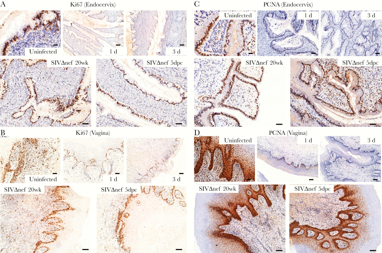
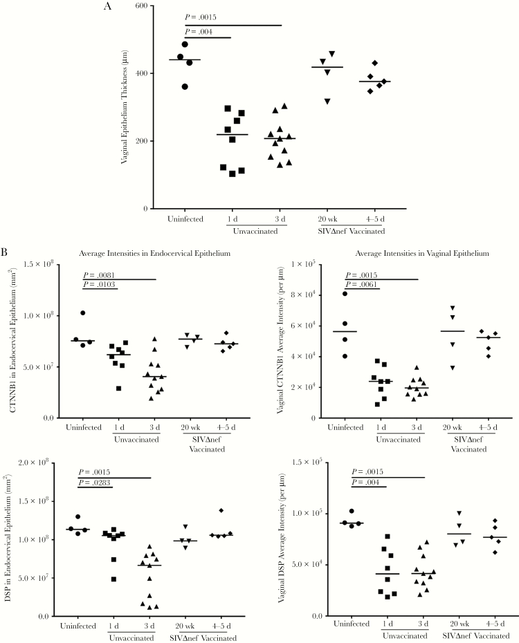
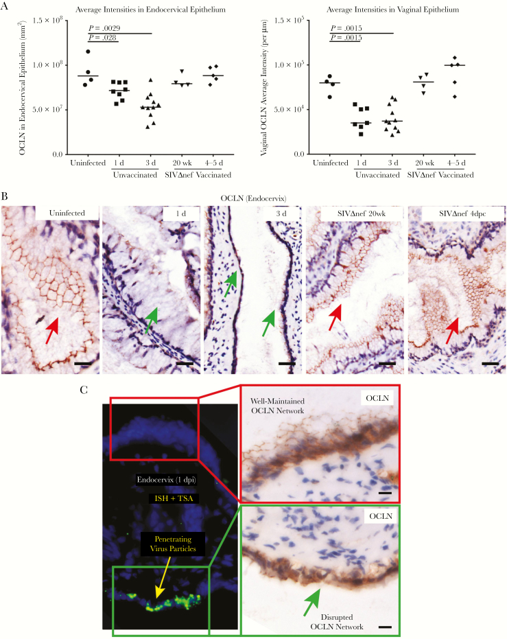
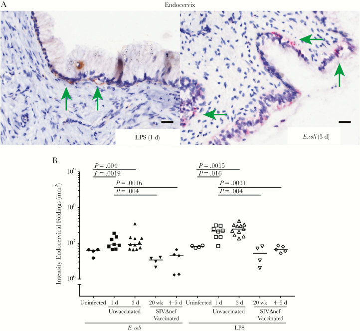
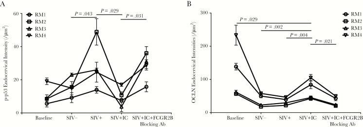
To identify the mechanisms by which human immunodeficiency virus type 1 (HIV-1) might penetrate the epithelial barrier during sexual transmission to women and the mechanisms of vaccine-associated protection against entry, we characterized early epithelial responses to vaginal inoculation of simian immunodeficiency virus strain mac251 (SIVmac251) in naive or SIVmac239Δnef-vaccinated rhesus macaques. Vaginal inoculation induced an early stress response in the cervicovaginal epithelium, which was associated with impaired epithelial integrity, damaged barrier function, and virus and bacterial translocation. In vaccinated animals, early stress responses were suppressed, and the maintenance of epithelial barrier integrity correlated with prevention of virus entry. These vaccine-protective effects were associated with a previously described mucosal system for locally producing and concentrating trimeric gp41 antibodies at the mucosal interface and with formation of SIV-specific immune complexes that block the stress responses via binding to the epithelial receptor FCGR2B and subsequent inhibitory signaling. Thus, blocking virus entry may be one protective mechanism by which locally concentrated non-neutralizing Ab might prevent HIV sexual transmission to women.


Figures

Figure 1.
Figure 1.
Vaginal inoculation induces cervical stress responses in unvaccinated rhesus macaques. A, Microarray analysis of the cervical tissues from uninfected and vaginally inoculated rhesus macaques (3–4 days after inoculation). The messenger RNA in the 1–2-mm surface layers of cervical tissues was extracted and subjected to microarray analysis. Genes were selected on the basis of their associated P values (<.05), false-positive cutoff q values (<0.2), and fold changes in expression (>1.2). Selected genes were then analyzed through Ingenuity Pathway analysis and grouped on the basis of their putative functions. Vaginal exposure to simian immunodeficiency virus (SIV) induced increased expression of genes related to a variety of stress responses. Fold changes in gene expression are indicated in the color scale bar. B, More than 50% of these stress response genes are associated with the DNA damage response and repair pathways. Genes were grouped on the basis of their known functions and associated signaling pathways. The percentages (x-axis) were calculated by dividing the number of genes in each group by the total number of genes. C, The densities of the p-p53–expressing nuclei in the endocervix significantly increased within 1 day after inoculation in unvaccinated animals but remained at baseline levels in SIVmac239Δnef-vaccinated animals before (20 weeks after vaccination) and after (4–5 days after challenge) vaginal challenge. Vaginal tissues of unvaccinated animals have a higher basal level of p-p53–expressing nuclei than the endocervix. The increases in p-p53–expressing nuclei were minimal 1 day after inoculation and then became significant 3 days after inoculation. However, in vaginal tissue specimens obtained from SIVmac239Δnef-vaccinated animals before and after vaginal challenge, the levels of p-p53–expressing nuclei were even lower than those at baseline. The nonparametric Wilcoxon signed rank test was used to compare the variations in expression of immunohistochemical markers across different groups over the course of infection. The bars represent median values in each group.
Figure 2.
Figure 2.
Vaginal inoculation induces stress responses in the cervicovaginal mucosa of unvaccinated rhesus macaques but not SIVmac239Δnef-vaccinated animals. Immunohistochemical staining demonstrates widespread increases in p-p53–expressing nuclei, mainly located in the epithelium in unvaccinated animals but not in vaccinated. A, In the endocervix, the p-p53–expressing nuclei were predominantly located in the endocervical epithelium after vaginal inoculation. B, In the vagina, p-p53–expressing nuclei were located in the basal layer of epithelium in uninfected animals and then quickly expanded into the adjoining epithelium and submucosa within 3 days after inoculation. In the SIVmac239Δnef-vaccinated animals, both the endocervical and vaginal epithelium were negative for p-p53 staining before and after vaginal challenge. All scale bars denote 50 µm.
Figure 3.
Figure 3.
Decreased proliferation and impaired epithelial homeostasis following vaginal inoculation in unvaccinated but not SIVmac239Δnef-vaccinated rhesus macaques. A and B, Immunohistochemical staining of the nuclear antigens Ki67 and PCNA were used as markers of proliferation. In uninfected animals, the Ki67-expressing nuclei were mainly located in the endocervical epithelium, the basal layers of vaginal epithelium, and the vaginal submucosa. Vaginal inoculation of simian immunodeficiency virus (SIV) was associated with dramatically reduced the numbers of positive nuclei to nearly undetectable levels in the endocervix and vaginal submucosa. In SIVmac239Δnef-vaccinated animals, the cervicovaginal epithelium still has strong expression of Ki67 after vaginal challenge. C and D, Similarly, the PCNA-expressing nuclei in the endocervix and vagina decreased quickly to nearly undetectable levels within 3 days after inoculation in unvaccinated animals but remained at a high level in SIVmac239Δnef-vaccinated animals before and after vaginal inoculation. The tissue distribution of PCNA-expressing nuclei is similar to that of Ki67-expressing nuclei, but PCNA was expressed in more nuclei than Ki67. All scale bars denote 50 µm.
Figure 4.
Figure 4.
Vaginal inoculation impairs the integrity of cervicovaginal epithelium in unvaccinated rhesus macaques but not vaccinated animals. A, Vaginal epithelium thinning within 1 day after inoculation in unvaccinated animals but not in SIVmac239Δnef-vaccinated animals after vaginal infection. First, the entire vaginal tissue section was scanned, and then the entire area of the vaginal epithelium and the length of the epithelium were quantified in ImageScope software. The average thickness of epithelial layer was acquired by dividing the epithelium area by the epithelium length. B, Decreased expression of β-catenin (CTNNB1) and desmoplakin (DSP) in the endocervix and vagina in unvaccinated animals but not in vaccinated animals after vaginal challenge. The nonparametric Wilcoxon signed rank test was used to compare the variations in expression of immunohistochemical markers across different groups over the course of infection. The bars represent median values in each group.
Figure 5.
Figure 5.
Virus penetration of damaged cervicovaginal epithelium in unvaccinated rhesus macaques but not vaccinated animals. A, Decreased expression of occludin (OCLN) in the endocervix and vagina of unvaccinated but not SIVmac239Δnef-vaccinated animals. The nonparametric Wilcoxon signed rank test was used to compare the variations in expression of immunohistochemical markers across different groups over the course of infection. The bars represent median values in each group. B, The OCLN network was continuous in uninfected and vaccinated animals in endocervical epithelium (red arrows), whereas, following vaginal inoculation, epithelial expression was discontinuous (green arrows) within 1 day after inoculation. Three days after inoculation, a continuous layer of OCLN-expressing endocervical epithelium was barely visible. The OCLN network was well maintained in the SIVmac239Δnef-vaccinated animals before and after vaginal challenge. C, The disruption of the OCLN network structure was spatially correlated with green virus particles and aggregates that penetrated the epithelial barrier. All scale bars denote 50 µm.
Figure 6.
Figure 6.
Increased bacterial translocation in unvaccinated but not vaccinated rhesus macaques. A, Immunohistochemical (IHC) staining with antibodies to lipopolysaccharide (LPS) and Escherichia coli detect bacteria underneath the columnar epithelium in the endocervix within 3 days after inoculation (green arrows; quantified in B), but not in the vaginal tissues. Bacteria and LPS were not detected in the submucosa of SIVmac239Δnef-vaccinated animals. All scale bars denote 50 µm. The nonparametric Wilcoxon signed rank test was used to compare the variations in expression of IHC markers across different groups over the course of infection. The bars represent median values in each group.
Figure 7.
Figure 7.
Immune complex-FCGR2B inhibitory signaling suppresses the stress responses to simian immunodeficiency virus (SIV) exposure in cervical tissues and maintains expression of cell-cell junction proteins in the epithelium. Pretreating fresh cervical explants of rhesus macaque ex vivo with SIV-specific immune complexes (ICs) before topical application of SIV suppressed the increases of p-p53–expressing nuclei (A) in the mucosa, and maintained to some degree the expression of occludin (OCLN; B) in the epithelium. These inhibitory effects were abrogated when the binding of ICs to FCGR2B was interrupted by blocking antibody (Ab). The nonparametric Wilcoxon signed rank test was used to compare the variations in expression of immunohistochemical markers across different treatment groups. The bars represent standard deviations among repeats in each group.

Similar articles

Cited by

References

    1. UN Joint Programme on HIV/AIDS (UNAIDS). Global Report: UNAIDS Report on the Global AIDS Epidemic. Geneva, Switzerland: UNAIDS, World Health Organization; 2014.
    1. Fauci AS. 25 years of HIV. Nature 2008; 453:289–90. - PubMed
    1. UN Joint Programme on HIV/AIDS (UNAIDS). Global Report: UNAIDS Report on the Global AIDS Epidemic. Geneva, Switzerland: UNAIDS, World Health Organization; 2013.
    1. Morison L. The global epidemiology of HIV/AIDS. Br Med Bull 2001; 58:7–18. - PubMed
    1. Quinn TC, Overbaugh J. HIV/AIDS in women: an expanding epidemic. Science 2005; 308:1582–3. - PubMed

Publication types

MeSH terms