. 2018 Feb 28;140(8):2841-2852.
doi: 10.1021/jacs.7b11811. Epub 2018 Feb 16.

Metabolomic "Dark Matter" Dependent on Peroxisomal β-Oxidation in Caenorhabditis elegans

Affiliations

Metabolomic "Dark Matter" Dependent on Peroxisomal β-Oxidation in Caenorhabditis elegans

Alexander B Artyukhin et al. J Am Chem Soc. .

Abstract

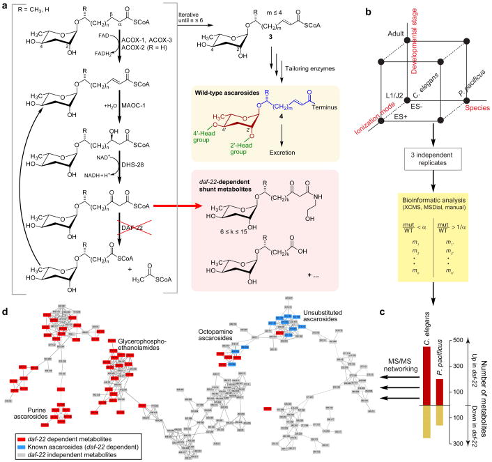
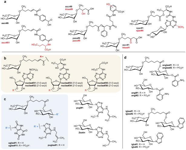
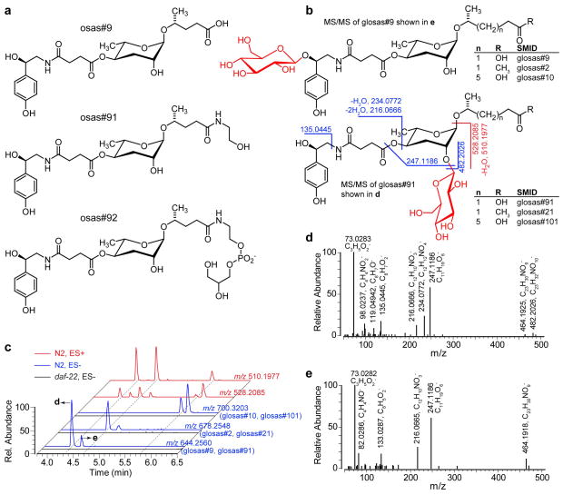
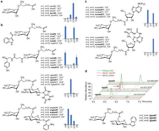
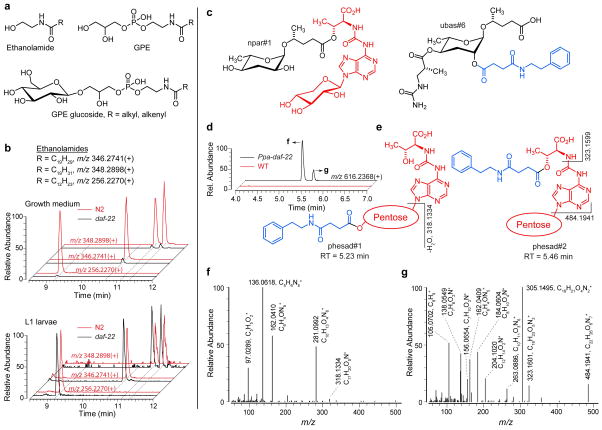
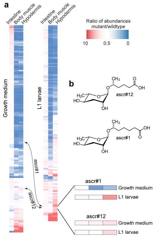
Peroxisomal β-oxidation (pβo) is a highly conserved fat metabolism pathway involved in the biosynthesis of diverse signaling molecules in animals and plants. In Caenorhabditis elegans, pβo is required for the biosynthesis of the ascarosides, signaling molecules that control development, lifespan, and behavior in this model organism. Via comparative mass spectrometric analysis of pβo mutants and wildtype, we show that pβo in C. elegans and the satellite model P. pacificus contributes to life stage-specific biosynthesis of several hundred previously unknown metabolites. The pβo-dependent portion of the metabolome is unexpectedly diverse, e.g., intersecting with nucleoside and neurotransmitter metabolism. Cell type-specific restoration of pβo in pβo-defective mutants further revealed that pβo-dependent submetabolomes differ between tissues. These results suggest that interactions of fat, nucleoside, and other primary metabolism pathways can generate structural diversity reminiscent of that arising from combinatorial strategies in microbial natural product biosynthesis.


Figures

Figure 1
Figure 1
Characterization of the daf-22-dependent portion of the C. elegans and P. pacificus metabolomes. a) Model for the biosynthesis of ascarosides. Iterative 4-step β-oxidation of long-chain ascarosides produces short-chain precursors of modular ascarosides that further functionalized at the carboxy terminus or at positions 2′ and/or 4′. daf-22 loss-of-function results in build-up of shunt metabolites, including long chain ascarosides and their ethanolamide derivatives. b) Schematic of experimental approach and analysis pipeline to detect and identify differential metabolites. Two species at two different developmental stages were analyzed in ESI+ and ESI− mode, resulting in 8 data sets (represented by black dots) c) Total number of daf-22-dependent metabolites in C. elegans and P. pacificus. d) Partial representation of MS/MS network (ES−) showing known ascarosides and newly discovered daf-22-dependent metabolites as part of diverse networks that represent a wide range of different metabolite families. See Supporting Information for full networks (Figure S2 and S3).
Figure 2
Figure 2
a) Examples of new ascaroside derivatives closely related to previously described compounds from C. elegans (ascr#9, ascr#8, ascr#1) and P. pacificus (npar#1, pasa#9). Known structures are in black, predicted new modifications are shown in red. The structure of ascr#81 was confirmed by total synthesis. b) Structures of ascarosylated ribonucleosides. Nuclas#31–36 occur as interconverting mixtures of the 2-O- and 3-O-ascarosylated isomers. c) Ascarosylated gluconucleosides uglas#1 and puglas#1, the highly abundant pugl#1, and the plant cytokinin zeatin. c) Anthranilic acid and indole derivative anglas#1 and iglas#1. Also shown are the previously described angl#1 and iglu#1, which are not daf-22-dependent.
Figure 3
Figure 3
a) Structures of osas#9 and examples of newly identified derivatives. b) Structures of two series of osas glucoside isomers with selected MS/MS fragmentations in ES− (blue) and ES+ (red) that distinguish isomers. c) EICs of osas glucosides. EICs in ES− (blue) correspond to glucosides of osas#9 (m/z 644.2560), osas#2 (m/z 678.2548) and osas#10 (m/z 700.3203). Neutral loss of fatty acid side chain in ES+ results in fragments common to all three osas glucosides (m/z 528.2085 for all glucosides and m/z 510.1977 after additional water loss, which is favored when the octopamine OH group is unsubstituted). d, e) MS/MS spectra of osas#9 glucosides in ES−. Absence of ions with m/z 135.0445, 234.0772, 482.2026 in e) suggests glycosylation of the octopamine hydroxy group in this isomer.
Figure 4
Figure 4
a) Relative abundances as determined by LC-MS of unsubstituted ascarosides. b) Relative abundances of selected families of 4′-modified ascarosides and ascaroside-glucoside combinations. c) Relative abundances of two families of ascarosylated nucleosides. d) LC-MS analysis of ascr#1 feeding experiment. EICs (ES−) of iglas#7 (m/z 570.2112), iglas#1 (m/z 572.2273) and iglas#3 (m/z 598.2424). These compounds are present in wildtype (N2) C. elegans (green) but absent in daf-22 medium (black). When synthetic ascr#1 is added to daf-22 cultures, the worms produce the ascr#1-derivative iglas#1 (red arrow) but not the corresponding ascr#7 or ascr#3 derivatives (red).
Figure 5
Figure 5
a) Generic structures of N-acylethanolamine, GPE, and GPE glucosides. b) Levels of EPEA (m/z 346.2741), arachidonic acid ethanolamides (AEA, m/z 348.2898) and other ethanolamides are significantly reduced in the metabolome of daf-22 growth medium, but largely unaffected in the L1 metabolome. The two peaks for AEA correspond to N- and O-acylethanolamines as determined by MS/MS (Tables S2–S9). c) Structures of npar#1 and ubas#6 previously identified from wildtype P pacificus. d) ES+ EICs for m/z 616.2368, showing two peaks present in Ppa-daf-22 but not in wildtype. Proposed structures for two isomers along with selected MS/MS fragmentations used in structure assignments are shown in e) and full ES+ MS/MS spectra for both isomers are shown in f) for phesad#1 and in g) for phesad#2.
Figure 6
Figure 6
Tissue specific daf-22 rescue in C. elegans (also see Figure S8). a) Heat maps showing expression of daf-22-dependent metabolites in tissue specific rescue lines relative to N2 levels in growth medium and L1 metabolomes (ES MS). b) Enlargement showing ascr#12 and ascr#1 expression patterns. In contrast to most other metabolites, daf-22 expression in any tissue rescues ascr#12 to near wildtype levels. ascr#1 can be effectively synthesized in the hypodermis and body muscle in L1 larvae but not in adults.

References

    1. Lodhi IJ, Semenkovich CF. Cell Metab. 2014;19(3):380–92. - PMC - PubMed
    1. Wanders RJA, Waterham HR. Ann Rev Biochem. 2006;75(1):295–332. - PubMed
    1. Nyathi Y, Baker A. Biochim Biophys Acta. 2006;1763(12):1478–95. - PubMed
    1. Baker A, Graham IA, Holdsworth M, Smith SM, Theodoulou FL. Trends Plant Sci. 2006;11(3):124–32. - PubMed
    1. von Reuss SH, Schroeder FC. Nat Prod Rep. 2015;32(7):994–1006. - PMC - PubMed

Publication types

LinkOut - more resources