. 2018 May;32(5):2855-2865.
doi: 10.1096/fj.201600788RR. Epub 2018 Jan 17.

Sphingosine-1-phosphate receptor 1 mediates elevated IL-6 signaling to promote chronic inflammation and multitissue damage in sickle cell disease

Affiliations

Sphingosine-1-phosphate receptor 1 mediates elevated IL-6 signaling to promote chronic inflammation and multitissue damage in sickle cell disease

Shushan Zhao et al. FASEB J. 2018 May.

Abstract

Sphingosine-1-phosphate (S1P) is a biolipid involved in chronic inflammation in several inflammatory disorders. Recent studies revealed that elevated S1P contributes to sickling in sickle cell disease (SCD), a devastating hemolytic, genetic disorder associated with severe chronic inflammation and tissue damage. We evaluated the effect of elevated S1P in chronic inflammation and tissue damage in SCD and underlying mechanisms. First, we demonstrated that interfering with S1P receptor signaling by FTY720, a U.S. Food and Drug Administration-approved drug, significantly reduced systemic, local inflammation and tissue damage without antisickling effects. These findings led us to discover that S1P receptor activation leads to substantial elevated local and systemic IL-6 levels in SCD mice. Genetic deletion of IL-6 in SCD mice significantly reduced local and systemic inflammation, tissue damage, and kidney dysfunction. At the cellular level, we determined that elevated IL-6 is a key cytokine functioning downstream of elevated S1P, which contributes to increased S1P receptor 1 ( S1pr1) gene expression in the macrophages of several tissues in SCD mice. Mechanistically, we revealed that S1P-S1PR1 signaling reciprocally up-regulated IL-6 gene expression in primary mouse macrophages in a JAK2-dependent manner. Altogether, we revealed that elevated S1P, coupled with macrophage S1PR1 reciprocally inducing IL-6 expression, is a key signaling network functioning as a malicious, positive, feed-forward loop to sustain inflammation and promote tissue damage in SCD. Our findings immediately highlight novel therapeutic possibilities.-Zhao, S., Adebiyi, M. G., Zhang, Y., Couturier, J. P., Fan, X., Zhang, H., Kellems, R. E., Lewis, D. E., Xia, Y. Sphingosine-1-phosphate receptor 1 mediates elevated IL-6 signaling to promote chronic inflammation and multitissue damage in sickle cell disease.

Keywords: JAK2; S1PR1; macrophage.


Conflict of interest statement

This work was supported by U.S. National Institutes of Health, National Heart, Lung, and Blood Institute Grants HL113574, HL114457, HL136969, and HL137990 (to Y.X.). The authors declare no conflicts of interest.

Figures
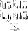
Figure 1.
Figure 1.
Anti-inflammatory effects of FTY720 treatment in SCD mice. SCD mice were treated with saline or FTY720. After 8 wk of treatment, the following parameters were measured. A) S1PR1 expression on CD3+CD4CD8 (double-negative) T-cells from lymph nodes and thymus of WT and SCD mice treated with FTY720 or saline. B) FTY720 treatment reduced plasma cytokine levels in SCD mice. OD, optical density. C) FTY720 treatment reduced plasma IL-6 levels in SCD mice. D) FTY720 treatment reduced IL-6 expression levels in spleen, liver, lung, and kidney of SCD mice. E) No effect of FTY720 on red blood cell life span in SCD mice. Values shown represent means ± sem (n = 6–7/group). *P < 0.01, WT mice treated with saline compared with WT mice treated with FTY720; **P < 0.05, SCD mice treated with saline compared with WT mice treated with saline or SCD mice treated with FTY720 compared with SCD mice treated with saline, respectively; #P < 0.001, SCD mice treated with FTY720 compared with SCD mice treated with saline.
Figure 2.
Figure 2.
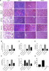
FTY720 treatment or genetic deletion of Il6 reduces multitissue damage and kidney dysfunction in SCD mice. A) H&E staining of lung, spleen, liver, and kidney. Arrowheads denote representative vascular congestion in lung and to necrosis in spleen, liver, and kidney. Scale bars, 200 μm. BE) Semiquantification of congestion or necrosis in lung, spleen, liver, and renal medulla. F) Ratio of microalbumin to creatinine in urine. G) Plasma IL-6 was abolished in SCD/Il6/ mice. *P < 0.01, SCD mice compared with WT mice; **P < 0.05, SCD mice treated with FTY720 compared with SCD mice treated with saline or SCD mice compared with SCD/Il6/ mice. ND, not determined.
Figure 3.
Figure 3.
FTY720 treatment and genetic deletion of Il6 reduce elevated S1PR1 expression in tissue macrophages of SCD mice. A, B) Expression of S1PR1 in F4/80+ macrophages of spleen (A) and kidney (B) of WT and SCD mice treated with saline or FTY720. Data are expressed as means ± sem (n = 6–8/group).*P < 0.05, WT mice treated with FTY720 vs. saline-treated WT mice; **P < 0.05, SCD mice treated with saline vs. saline-treated WT mice; #P < 0.05, FTY720-treated SCD mice vs. saline treated SCD mice. C, D) Expression of S1PR1 in F4/80+ macrophages of spleen (C) and kidney (D) of control, SCD, and SCD/Il6/ mice. Data are expressed as means ± sem (n = 6–8/group). *P < 0.05, SCD mice compared with WT control mice; **P < 0.05, SCD mice compared with SCD/Il6/ mice.
Figure 4.
Figure 4.
S1P signaling via S1PR1 directly induces Il6 gene expression and elevated Il6 reciprocally up-regulates S1PR1 gene expression in a JAK2-dependent manner in primary, cultured mouse macrophages. Mouse bone marrow from WT and Il6−/− mice were isolated and differentiated to macrophages following with treatment of S1P in the presence or absence of S1P receptor specific antagonists or JAK2 inhibitor. A, B) Il6 gene expression (A) and protein level (B) in the medium of cultured WT mouse primary macrophages. C) S1pr1 mRNA levels in WT and Il6−/− cultured primary macrophages. D–F) Il6 gene expression (D); secreted IL-6 protein levels (E); and S1pr1 gene expression (F) in cultured WT mouse primary macrophages with treatment of DMSO, S1P, or AG490 (a JAK2 inhibitor). Data are expressed as means ± sem (n = 6/group). *P < 0.01, S1P treated group compared with DMSO-treated group; **P < 0.05, S1P-treated group compared with FTY720-treated group, S1P-treated group compared with W146-treated group, and S1P-treated group compared with AG490-treated group. G) Working model of elevated S1P with dual consequences in SCD: 1) it functions intracellularly within erythrocytes to induce sickling (22); and 2) it functions extracellularly via S1PR1 signaling in macrophages to induce chronic inflammation and tissue damage. Elevated plasma S1P activating via S1PR1 on macrophages up-regulates Il6 gene expression and production in a JAK2-dependent manner that reciprocally induce S1pr1 gene expression. Treatment with FTY720 inhibits S1PR1 activation, or genetic deletion of Il6 successfully reduced sustained inflammation and tissue damage in SCD mice.

References

    1. Maceyka M., Harikumar K. B., Milstien S., Spiegel S. (2012) Sphingosine-1-phosphate signaling and its role in disease. Trends Cell Biol. 22, 50–60 10.1016/j.tcb.2011.09.003 - DOI - PMC - PubMed
    1. An S., Zheng Y., Bleu T. (2000) Sphingosine 1-phosphate-induced cell proliferation, survival, and related signaling events mediated by G protein-coupled receptors Edg3 and Edg5. J. Biol. Chem. 275, 288–296 10.1074/jbc.275.1.288 - DOI - PubMed
    1. Garcia J. G., Liu F., Verin A. D., Birukova A., Dechert M. A., Gerthoffer W. T., Bamberg J. R., English D. (2001) Sphingosine 1-phosphate promotes endothelial cell barrier integrity by Edg-dependent cytoskeletal rearrangement. J. Clin. Invest. 108, 689–701 10.1172/JCI12450 - DOI - PMC - PubMed
    1. Lee O. H., Kim Y. M., Lee Y. M., Moon E. J., Lee D. J., Kim J. H., Kim K. W., Kwon Y. G. (1999) Sphingosine 1-phosphate induces angiogenesis: its angiogenic action and signaling mechanism in human umbilical vein endothelial cells. Biochem. Biophys. Res. Commun. 264, 743–750 10.1006/bbrc.1999.1586 - DOI - PubMed
    1. Liang J., Nagahashi M., Kim E. Y., Harikumar K. B., Yamada A., Huang W. C., Hait N. C., Allegood J. C., Price M. M., Avni D., Takabe K., Kordula T., Milstien S., Spiegel S. (2013) Sphingosine-1-phosphate links persistent STAT3 activation, chronic intestinal inflammation, and development of colitis-associated cancer. Cancer Cell 23, 107–120 10.1016/j.ccr.2012.11.013 - DOI - PMC - PubMed

Publication types

MeSH terms

LinkOut - more resources