. 2018 Jun;32(6):3385-3397.
doi: 10.1096/fj.201701145R. Epub 2018 Jan 22.

Differential susceptibility of Dectin-1 isoforms to functional inactivation by neutrophil and fungal proteases

Affiliations

Differential susceptibility of Dectin-1 isoforms to functional inactivation by neutrophil and fungal proteases

James S Griffiths et al. FASEB J. 2018 Jun.

Abstract

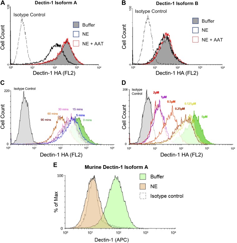
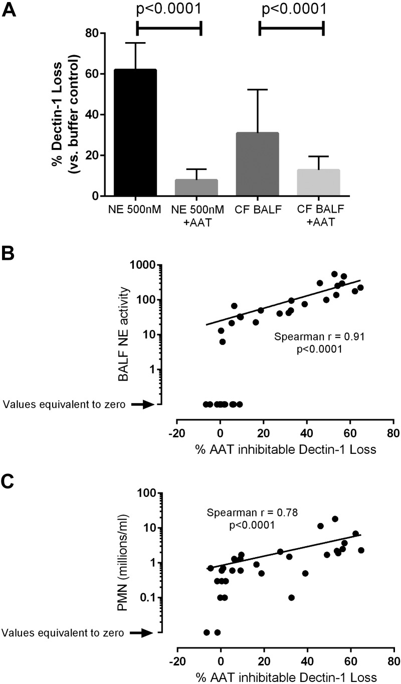
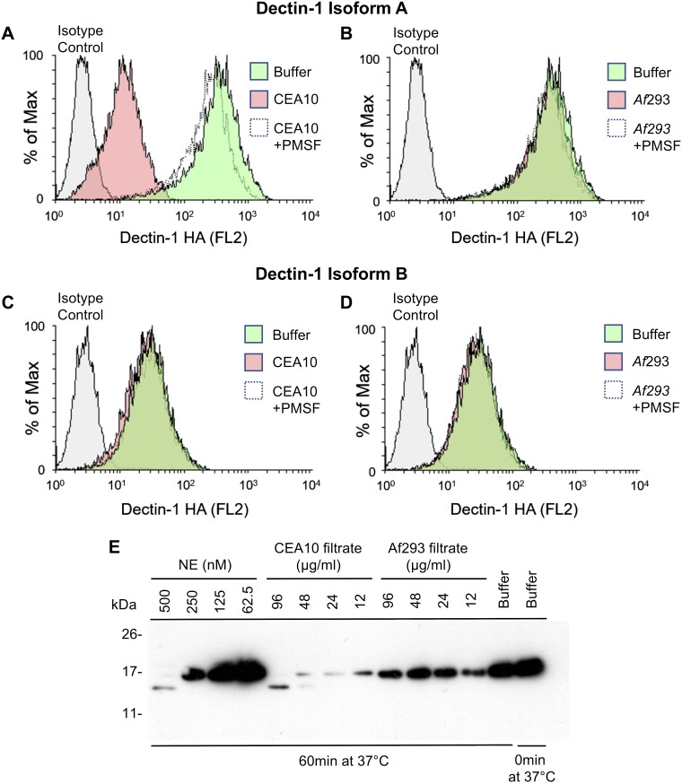
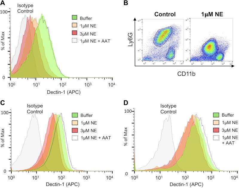
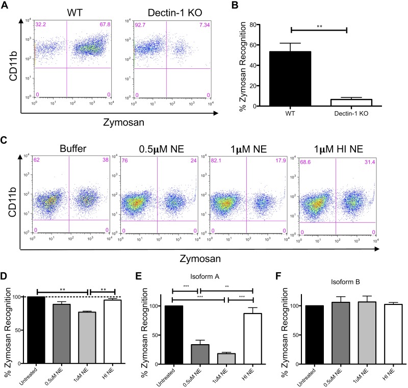
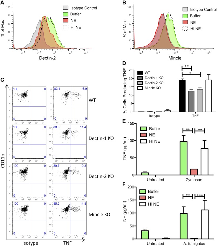
Patients with cystic fibrosis (CF) experience chronic or recurrent bacterial and fungal lung infections. Many patients with CF cannot effectively clear Aspergillus from their lungs. This may result in IgE sensitization and the development of allergic bronchopulmonary aspergillosis, or invasive infections, such as Aspergillus bronchitis. Lung disease in patients with CF is associated with neutrophil-dominated inflammation and elevated levels of the serine protease, neutrophil elastase (NE). Various C-type lectin-like receptors (CLRs), including Dectin-1 and Dectin-2, are involved in the immune response to Aspergillus. Here, we show that purified NE cleaves Dectin-1 in an isoform-specific manner. Bronchoalveolar lavage fluid from patients with CF, which contains high NE activity, induces Dectin-1 cleavage. Similarly, filtrate from a protease-producing strain of Aspergillus fumigatus induces isoform-specific cleavage of Dectin-1. Dectin-1 knockout (KO) cells and NE-treated cells demonstrated reduced phagocytosis of zymosan, a fungal cell wall preparation. In addition, NE cleaves 2 other CLRs, Dectin-2 and Mincle, and fungal-induced cytokine production was reduced in Dectin-1 KO cells, Dectin-2 KO cells, and NE-treated cells. Thus, Dectin-1 and Dectin-2 cleavage by NE and/or A. fumigatus-derived proteases results in an aberrant antifungal immune response that likely contributes to disease pathology in patients with CF.-Griffiths, J. S., Thompson, A., Stott, M., Benny, A., Lewis, N. A., Taylor, P. R., Forton, J., Herrick, S., Orr, S. J., McGreal, E. P. Differential susceptibility of Dectin-1 isoforms to functional inactivation by neutrophil and fungal proteases.

Keywords: Aspergillus; C-type lectin-like receptor; cystic fibrosis; elastase.


Figures

Figure 1.
Figure 1.
Dectin-1A is cleaved by NE. AC) NIH-3T3 cells that express human HA-tagged Dectin-1 isoform A (A, C) or Dectin-1 isoform B (B) were exposed to 0.5 µM NE, in the presence or absence of AAT, for 30 min (A, B) or for the indicated times (C) at 37°C. Cells were stained with an anti-HA Ab and analyzed by flow cytometry. D) NIH-3T3 cells that express human HA-tagged Dectin-1 isoform A were exposed to the indicated concentrations of NE for 30 min at 37°C. Cells were stained with an anti-HA Ab and analyzed by flow cytometry. E) NIH-3T3 cells that express murine Dectin-1 isoform A were exposed to NE for 1 h at 37°C. Cells were stained with an anti–Dectin-1 Ab and analyzed by flow cytometry. Data are representative of 3–4 independent experiments. APC, allophycocyanin.
Figure 2.
Figure 2.
CF BALF induces cleavage of Dectin-1. A) NIH-3T3 cells that express human HA-tagged Dectin-1 isoform A were exposed to 0.5 µM NE or 50 µl CF BALF, in the presence or absence of AAT, for 30 min at 37°C. Cells were stained with anti-HA Ab and analyzed by flow cytometry. Graph displays means ± sd. Data are the cumulative result of 7 independent experiments. B, C) NIH3T3 cells that express human HA-tagged Dectin-1 isoform A were exposed to CF BALF for 30 min at 37°C. Cells were stained with an anti-HA Ab and analyzed by flow cytometry. NE activity in CF BALF samples was measured (B). Correlation analysis between NE activity and percentage loss of Dectin-1 was performed. Neutrophil numbers in the CF BALF were counted and correlation analysis between neutrophil numbers in CF BALF samples and percentage loss of Dectin-1 was performed (C). Graphs are the cumulative result of at least 7 independent experiments (B, C). Each symbol represents data for CF BALF from a single patient.
Figure 3.
Figure 3.
A. fumigatus–derived proteases cleave Dectin-1. AD) NIH3T3 cells that express human HA-tagged Dectin-1 isoform A (A, B) or isoform B (C, D) were exposed to A. fumigatus CEA10 supernatant (A, C) or Af293 supernatant (B, D)—in the presence or absence of PMSF—for 30 min at 37°C. Cells were stained with anti-HA Ab and analyzed by flow cytometry. Plots are representative of 3 independent experiments. E) Soluble Dectin-1 was exposed to NE or A. fumigatus filtrates from protease-producing (CEA10) or non–protease-producing (Af293) fungal strains at the indicated concentrations for 60 min at 37°C. Proteases were inhibited with PMSF, and Dectin-1 was separated by SDS-PAGE, blotted to nitrocellulose membrane, and probed with anti–Dectin-1. Data are representative of 3 independent experiments.
Figure 4.
Figure 4.
NE cleaves Dectin-1 on myeloid cells. A) BMDMs from BALB/c mice were exposed to NE, in the presence or absence of AAT, for 1 h at 37°C. Cells were stained with anti–Dectin-1 and analyzed by flow cytometry. BD) BALB/c mice were injected i.p. with 0.5 ml Biogel, and inflammatory infiltrates were recovered 16 h later by peritoneal lavage. Cells were left unexposed or exposed to NE in the presence or absence of AAT for 1 h at 37°C. Cells were stained with anti-Ly6G, anti-CD11b, and anti–Dectin-1, and analyzed by flow cytometry. Dectin-1 levels were measured in the LyG+CD11b+ (neutrophil) population (C) and LyGCD11b+ (inflammatory monocyte/macrophage) population (D). Data are representative of 3 independent experiments.
Figure 5.
Figure 5.
NE cleavage of Dectin-1 impairs zymosan recognition. AD) Wild-type (WT) and Dectin-1 KO BMDMs (A, B) and NE-treated BMDMs from BALB/c mice (C, D) were incubated with 2.5 µg FITC-labeled zymosan for 15 min. Cells were stained with anti-F4/80, anti-CD11b, and anti–Dectin-1, and analyzed by flow cytometry. Data are representative of 3 independent experiments. Zymosan recognition in Dectin-1 KO cells (B) and NE/HI NE-treated cells (D) is relative to WT cells (set at 100%; B) or buffer control cells (set at 100%; D). E, F) NE-treated NIH3T3 cells that express hDectin-1 isoform A (E) or isoform B (F) were incubated with 2.5 µg FITC-labeled zymosan for 15 min. Cells were stained with anti–Dectin-1 and analyzed by flow cytometry. Data are representative of 3 independent experiments. Zymosan recognition in NE/HI NE-treated cells is relative to buffer control cells (set at 100%). Graphs display means ± sem. Representative data from 1 of the 3 independent experiments (A, C). **P < 0.01, Student’s t test (B); **P < 0.01, ***P < 0.001, 1-way ANOVA with Bonferroni’s posttest (D, E).
Figure 6.
Figure 6.
NE cleaves CLRs and reduces TNF production. A, B) BALB/c mice were injected with Biogel (0.5 ml, i.p.), and inflammatory infiltrates were recovered 16 h later by peritoneal lavage. Cells were left unexposed or exposed to 1 µM NE or HI NE for 1 h at 37°C. Cells were stained with anti-Ly6G, anti-CD11b, anti–Dectin-2, and anti-Mincle, and analyzed by flow cytometry. Dectin-2 (A) and Mincle (B) levels were measured in the LyGCD11b+ inflammatory monocyte/macrophage population. C, D) Wild-type (WT), Dectin-1 KO, Dectin-2 KO, and Mincle KO mice were injected intraperitoneally with 0.5 ml Biogel, and inflammatory infiltrates were recovered 16 h later by peritoneal lavage. Cells were stimulated with cell trace far red-labeled A. fumigatus for 3 h in the presence of Brefeldin, and cells were stained with anti-Ly6G, anti-CD11b, anti-CD19, and anti-TNF, and analyzed by flow cytometry. Percentages of TNF-producing inflammatory monocytes with bound/phagocytosed A. fumigatus were measured. Representative data from 1 of 3 independent experiments is shown in panel C. Graph displays means ± sem (D). Graph displays cumulative data from 3 independent experiments. *P < 0.05, **P < 0.01, 2-way ANOVA with Bonferroni’s posttest. E) BALB/c mice were injected with Biogel (0.5 ml, i.p.), and inflammatory infiltrates were recovered 16 h later by peritoneal lavage. Cells were left unexposed or exposed to 2 µM NE or HI NE for 1 h at 37°C, followed by stimulation with 25 µg/ml zymosan for 4 h. TNF levels in the supernatants were measured by ELISA. Graph displays means ± sem. Graph displays cumulative data from 3 independent experiments. **P < 0.01, 2-way ANOVA with Bonferroni’s posttest. F) BALB/c mice were injected with Biogel (0.5 ml, i.p.), and inflammatory infiltrates were recovered 16 h later by peritoneal lavage. The inflammatory monocyte population was enriched by Ly6G+ cell depletion. Cells were left unexposed or exposed to 2 µM NE or HI NE for 1 h at 37°C, followed by stimulation with A. fumigatus for 4 h. TNF levels in the supernatants were measured by ELISA. Graph displays means ± sem. Graph displays cumulative data from 4 independent experiments. **P < 0.01, ***P < 0.001, paired 2-way ANOVA with Bonferroni’s posttest.

References

    1. Brown G. D., Denning D. W., Gow N. A., Levitz S. M., Netea M. G., White T. C. (2012) Hidden killers: human fungal infections. Sci. Transl. Med. 4, 165rv13 - PubMed
    1. King J., Brunel S. F., Warris A. (2016) Aspergillus infections in cystic fibrosis. J. Infect. 72(Suppl), S50–S55 - PubMed
    1. El-Dahr J. M., Fink R., Selden R., Arruda L. K., Platts-Mills T. A., Heymann P. W. (1994) Development of immune responses to Aspergillus at an early age in children with cystic fibrosis. Am. J. Respir. Crit. Care Med. 150, 1513–1518 - PubMed
    1. Baxter C. G., Dunn G., Jones A. M., Webb K., Gore R., Richardson M. D., Denning D. W. (2013) Novel immunologic classification of aspergillosis in adult cystic fibrosis. J. Allergy Clin. Immunol. 132, 560–566.e510 - PubMed
    1. Plato A., Hardison S. E., Brown G. D. (2015) Pattern recognition receptors in antifungal immunity. Semin. Immunopathol. 37, 97–106 - PMC - PubMed

Publication types

MeSH terms

LinkOut - more resources