Review
. 2018 Feb 3;23(2):323.
doi: 10.3390/molecules23020323.

Recent Advances in Detecting Mitochondrial DNA Heteroplasmic Variations

Affiliations
Review

Recent Advances in Detecting Mitochondrial DNA Heteroplasmic Variations

Mengqin Duan et al. Molecules. .

Abstract

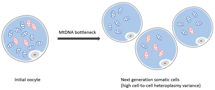
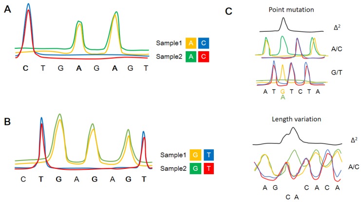
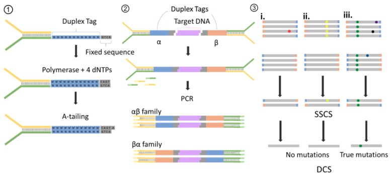
The co-existence of wild-type and mutated mitochondrial DNA (mtDNA) molecules termed heteroplasmy becomes a research hot point of mitochondria. In this review, we listed several methods of mtDNA heteroplasmy research, including the enrichment of mtDNA and the way of calling heteroplasmic variations. At the present, while calling the novel ultra-low level heteroplasmy, high-throughput sequencing method is dominant while the detection limit of recorded mutations is accurate to 0.01% using the other quantitative approaches. In the future, the studies of mtDNA heteroplasmy may pay more attention to the single-cell level and focus on the linkage of mutations.

Keywords: heteroplasmy; mitochondria; mtDNA; next generation sequencing.


Conflict of interest statement

The authors declare no conflict of interest.

Figures

Figure 1
Figure 1
Bottleneck effect. There are two types of mtDNA in initial oocyte (left), where blue stand for mitochondria harboring wild-type mtDNA and red ones represent mutated ones. During the meiosis acting under the mtDNA bottleneck effect, the mitochondria in next generation somatic cells take on a high cell-to-cell heteroplasmy variance situation (right). Some of them may harbor all wild-type mtDNA while others accumulate one or more mutations.
Figure 2
Figure 2
A brief schematic diagram of enriching methods for mtDNA. Isolation methods separate or capture mtDNA from specimen directly, which need abounding biospecimen. Amplifying mtDNA methods whose mainly approaches are PCR and WGA, make the requirement for amount and freshness of biospecimen more broaden, and mtDNA can be amplified specifically. Bioinformatics methods can dig mtDNA data from other relevant genome sequencing data like exome and whole genome in cells.
Figure 3
Figure 3
A schematic diagram of polymorphism ratio sequencing (PRS). (A,B) present the four-color labeling scheme for A/C and G/T in different samples. (C) shows the Δ2 plot generated by squaring the differences between two samples, including the detection of point mutations and short length variations . The resolution of Δ2 plot (peak height) is about 5%.
Figure 4
Figure 4
The design of primers in ARMS-qPCR. A set of primers in ARMS is composed of 3 primers, including one common revers primer (blue) and two allele-specific (AS) forward primers (orange and green). The last base of AS primers are set purposively to be consistent with altered bases, while the two nucleotides immediately 5’ to the mutated site (marked as red) are also changed to increase the resolving power of ARMS.
Figure 5
Figure 5
BEAMing procedure. Step 1: Magnetic beads coated with streptavidin are bound to biotinylated oligonucleotides (oligos). Step 2: An aqueous mix containing all the necessary components of PCR, beads and template DNA are stirred together with an oildetergent mix to create microemulsions. Red and green lines represent two kinds of templates which may differ from each other by one or more nucleotides. The aqueous compartments (blue circles in grey oil layer) are fully diluted to ensure each of them contain an average of less than one template molecule and less than one bead. Step 3: These microemulsions are subjected to thermal cycles of conventional PCR. If a DNA template and a bead are present to be together in a single aqueous compartment, the bead-bound oligos act as a primer for amplification. Step 4: Breaking the emulsions and the beads are purified with a magnet. Step 5: Fluorescently labeled primers are hybridized to the amplified target DNA molecules, which renders the beads as specific signal after appropriate laser excitation. Step 6: Flow cytometry is used to count the beads with different fluorescence signals.
Figure 6
Figure 6
Procedure of duplex sequencing (DS). (1) Adapter synthesis. A 12 randomized base Duplex Tag is appended to one of two sequencing adapters, and then complete the complementary strand with DNA polymerase. Complete adapter A-tailing is ensured by extended incubation with polymerase and dATP. (2) Duplex sequencing working flow. Shared, T-tailed target double-stranded DNA molecule is ligated to A-tailed adapters. After PCR amplified with primers of adapter, two types of attachment are produced. Those derived from one target DNA fragment will have the α tag sequence adjacent to flow-cell adapter sequence 1 (yellow) and the β tag adjacent to flow-cell sequence 2 (green). (3) Error correction. Reads sharing a unique set of tags are grouped into paired families with members having strands labeled reciprocally (i–iii). (i) Mutation (red spot) present in only one or a few family members represent sequencing mistakes or PCR-introduced errors occurring late in amplification. (ii) Mutations (yellow spots) present in some or all the fragments of one family member but not in the other member. This situation represent that those mutation arise from PCR errors during the first round of amplification. (iii) When mutations occur on both strands of a DNA fragment appear in all members of a family pair, those are true mutations. Then reads with paired tags are grouped into single-stranded consensus sequences (SSCSs) with the bases of SSCSs fragments depend on the optimal base of each position. After that, SSCSs are reduced into duplex consensus sequences (DCSs).

References

    1. Anderson S., Bankier A.T., Barrell B.G., De Bruijn M., Coulson A.R., Drouin J., Eperon I., Nierlich D., Roe B.A., Sanger F. Sequence and organization of the human mitochondrial genome. Nature. 1981;290:457–465. doi: 10.1038/290457a0. - DOI - PubMed
    1. Chatterjee A., Mambo E., Sidransky D. Mitochondrial DNA mutations in human cancer. Oncogene. 2006;25:4663–4674. doi: 10.1038/sj.onc.1209604. - DOI - PubMed
    1. Wallace D.C., Singh G., Lott M.T., Hodge J.A., Schurr T.G., Lezza A.M.S., Elsas L.J., Nikoskelainen E. Mitochondrial DNA mutation associated with leber’s hereditary optic neuropathy. Science. 1988;242:1427–1430. doi: 10.1126/science.3201231. - DOI - PubMed
    1. Lightowlers R.N., Chinnery P.F., Turnbull D.M., Howell N. Mammalian mitochondrial genetics: Heredity, heteroplasmy and disease. Trends Genet. 1997;13:450–455. doi: 10.1016/S0168-9525(97)01266-3. - DOI - PubMed
    1. Naue J., Horer S., Sanger T., Strobl C., Hatzer-Grubwieser P., Parson W., Lutz-Bonengel S. Evidence for frequent and tissue-specific sequence heteroplasmy in human mitochondrial DNA. Mitochondrion. 2015;20:82–94. doi: 10.1016/j.mito.2014.12.002. - DOI - PubMed

MeSH terms

Substances