. 2018 Feb 5;10(2):43.
doi: 10.3390/cancers10020043.

Elevated Polyamines in Saliva of Pancreatic Cancer

Affiliations

Elevated Polyamines in Saliva of Pancreatic Cancer

Yasutsugu Asai et al. Cancers (Basel). .

Abstract

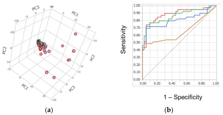
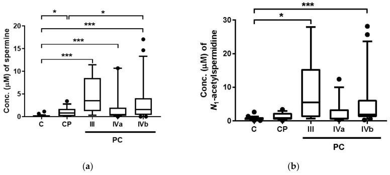
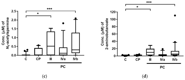
Detection of pancreatic cancer (PC) at a resectable stage is still difficult because of the lack of accurate detection tests. The development of accurate biomarkers in low or non-invasive biofluids is essential to enable frequent tests, which would help increase the opportunity of PC detection in early stages. Polyamines have been reported as possible biomarkers in urine and saliva samples in various cancers. Here, we analyzed salivary metabolites, including polyamines, using capillary electrophoresis-mass spectrometry. Salivary samples were collected from patients with PC (n = 39), those with chronic pancreatitis (CP, n = 14), and controls (C, n = 26). Polyamines, such as spermine, N₁-acetylspermidine, and N₁-acetylspermine, showed a significant difference between patients with PC and those with C, and the combination of four metabolites including N₁-acetylspermidine showed high accuracy in discriminating PC from the other two groups. These data show the potential of saliva as a source for tests screening for PC.

Keywords: metabolomics; pancreatic cancer; polyamines; saliva.


Conflict of interest statement

The authors declare no conflict of interest.

Figures

Figure 1
Figure 1
Discrimination ability of metabolomics profile: (a) Score plots of principal component analysis (PCA). Contribution ratio to first, second, and third principal component (PC) (PC1, PC2, and PC3) were 34.0, 5.7, and 4.7, respectively. Blue, green, and red plots indicated C, CP, and PC, respectively. (b) Receiver operating characteristic (ROC) curves of multiple logistic regression (MLR): red, green, blue, and orange curves indicated the MLR model with 4, 3, 2, and 1 metabolite(s), respectively, and their area under ROC curve (AUC) values were 0.887 (95% confidence interval (CI); 0.784–0.944, p < 0.0001), 0.859 (95% CI; 0.749–0.925, p < 0.0001), 0.807 (95% CI; 0.749–0.925, p < 0.0001), and 0.653 (95% CI; 0.526–0.761, p < 0.0122), respectively. The differences in AUC of the model with 4 parameters were 0.0501, 0.0280, and <0.0001 for those with 3, 2, and 1 parameters, respectively.
Figure 2
Figure 2
Salivary concentration of four metabolites showing significant difference among control patients (C), chronic pancreatitis patients (CP), and pancreatic cancer patients (PC): (a) spermine; (b) N1-acetylspermidine; (c) N1-acetylspermine; (d) 2-aminobutanoate (2AB). III, IVa, and IVb indicate the stage of PC. The number of subjects for C, CP, and PC Stages III, IVa, IVb were 26, 14, 6, 12, and 21, respectively. Horizontal bars of box-whisker plots indicate 10%, 90%, median, and lower and upper quantiles. The data <10% and >90% were depicted as plots. *** p <0.001 and * p < 0.05 by the Steel–Dwass test.
Figure 2
Figure 2
Salivary concentration of four metabolites showing significant difference among control patients (C), chronic pancreatitis patients (CP), and pancreatic cancer patients (PC): (a) spermine; (b) N1-acetylspermidine; (c) N1-acetylspermine; (d) 2-aminobutanoate (2AB). III, IVa, and IVb indicate the stage of PC. The number of subjects for C, CP, and PC Stages III, IVa, IVb were 26, 14, 6, 12, and 21, respectively. Horizontal bars of box-whisker plots indicate 10%, 90%, median, and lower and upper quantiles. The data <10% and >90% were depicted as plots. *** p <0.001 and * p < 0.05 by the Steel–Dwass test.

References

    1. Jemal A., Siegel R., Ward E., Hao Y., Xu J., Murray T., Thun M.J. Cancer statistics, 2008. CA Cancer J. Clin. 2008;58:71–96. doi: 10.3322/CA.2007.0010. - DOI - PubMed
    1. Rosty C., Goggins M. Early detection of pancreatic carcinoma. Hematol./Oncol. Clin. N. Am. 2002;16:37–52. doi: 10.1016/S0889-8588(01)00007-7. - DOI - PubMed
    1. Canto M.I., Hruban R.H., Fishman E.K., Kamel I.R., Schulick R., Zhang Z., Topazian M., Takahashi N., Fletcher J., Petersen G., et al. Frequent detection of pancreatic lesions in asymptomatic high-risk individuals. Gastroenterology. 2012;142:796–804. doi: 10.1053/j.gastro.2012.01.005. quiz e714-795. - DOI - PMC - PubMed
    1. Papadatos-Pastos D., Thillai K., Rabbie R., Ross P., Sarker D. Folfirinox—A new paradigm in the treatment of pancreatic cancer. Expert Rev. Anticancer Ther. 2014;14:1115–1125. doi: 10.1586/14737140.2014.957188. - DOI - PubMed
    1. Kiriyama S., Hayakawa T., Kondo T., Shibata T., Kitagawa M., Ono H., Sakai Y. Usefulness of a new tumor marker, span-1, for the diagnosis of pancreatic cancer. Cancer. 1990;65:1557–1561. doi: 10.1002/1097-0142(19900401)65:7<1557::AID-CNCR2820650718>3.0.CO;2-W. - DOI - PubMed

LinkOut - more resources