Review
. 2018 Feb 5;18(2):465.
doi: 10.3390/s18020465.

Intracranial Pressure Monitoring-Review and Avenues for Development

Affiliations
Review

Intracranial Pressure Monitoring-Review and Avenues for Development

Maya Harary et al. Sensors (Basel). .

Abstract

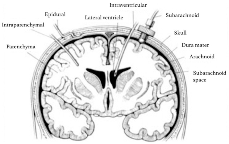
Intracranial pressure (ICP) monitoring is a staple of neurocritical care. The most commonly used current methods of monitoring in the acute setting include fluid-based systems, implantable transducers and Doppler ultrasonography. It is well established that management of elevated ICP is critical for clinical outcomes. However, numerous studies show that current methods of ICP monitoring cannot reliably define the limit of the brain's intrinsic compensatory capacity to manage increases in pressure, which would allow for proactive ICP management. Current work in the field hopes to address this gap by harnessing live-streaming ICP pressure-wave data and a multimodal integration with other physiologic measures. Additionally, there is continued development of non-invasive ICP monitoring methods for use in specific clinical scenarios.

Keywords: cerebral compliance; intracranial pressure monitoring; neurocritical care.


Conflict of interest statement

The authors declare no conflict of interest.

Figures

Figure 1
Figure 1
The Monro–Kellie model for the contents of the intracranial compartment. ‘Brain tissue’ includes neurons, glia, extracellular fluid and cerebral microvasculature. ‘Venous’ and ‘Arterial blood’ represents the intracranial blood volume in macro-vasculature and cerebral venous sinuses. ‘CSF’ includes ventricular and cisternal CSF.
Figure 2
Figure 2
Pressure–volume curve for ICP. The pressure–volume curve has four ‘zones’: (1) baseline intracranial volume with good compensatory reserve and high compliance (blue); (2) gradual depletion of compensatory reserve as intracranial volume increases (yellow); (3) poor compensatory reserve and increased risk of cerebral ischemia and herniation (red); and (4) critically high ICP causing collapse of cerebral microvasculature and disturbed cerebrovascular reactivity (grey).
Figure 3
Figure 3
Cerebral autoregulation capacity.
Figure 4
Figure 4
Sites for invasive ICP monitoring. These sites represent actual and potential spaces in the intracranial cavity in which ICP can be measured. Intraventricular monitoring with EVDs is the most commonly accessed site in clinical practice, followed by intraparenchymal probes. Reproduced with permission [32].
Figure 5
Figure 5
ICP pressure waves. (A) ICP fluctuations in response to the respiratory cycle (W2) and the arterial cycle (W1); (B) close-up of ICP waveform due to the systemic arterial cycle. Components are P1 (Percussion wave = representative of arterial pulsation), P2 (Tidal wave = a proxy for intracranial compliance) and P3 (Dicrotic wave = pressure transmission of aortic valve closure). A raised P2 wave is an indicator of raised ICP and reduced intracranial compliance (*); (C) Lundberg A (plateau) and B waves; adapted from Hall et al. [55].

References

    1. Monro A. Observations on the Structure and Functions of the Nervous System. Creech and Johnson; Edinbourgh, UK: 1783.
    1. Kellie G. Appearances observed in the dissection of two individuals; death from cold and congestion of the brain. Trans. Med.-Chir. Soc. Edinbrugh. 1824;1:84. - PMC - PubMed
    1. Cushing H. The Third Circulation in Studies in Intracranial Physiology and Surgery. Oxford University Press; London, UK: 1926.
    1. Greenberg M., editor. Handbook of Neurosurgery. 8th ed. Thieme; New York, NY, USA: 2016. Neuromonitoring; pp. 856–881.
    1. Leffert L.R., Schwamm L.H. Neuraxial anesthesia in parturients with intracranial pathology: A comprehensive review and reassessment of risk. Anesthesiology. 2013;119:703–718. doi: 10.1097/ALN.0b013e31829374c2. - DOI - PubMed

LinkOut - more resources