

Trifunctional High-Throughput Screen Identifies Promising Scaffold To Inhibit Grp94 and Treat Myocilin-Associated Glaucoma
- PMID: 29402077
- PMCID: PMC6195314
- DOI: 10.1021/acschembio.7b01083
Trifunctional High-Throughput Screen Identifies Promising Scaffold To Inhibit Grp94 and Treat Myocilin-Associated Glaucoma
Abstract
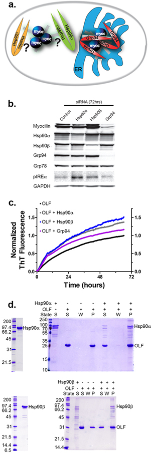
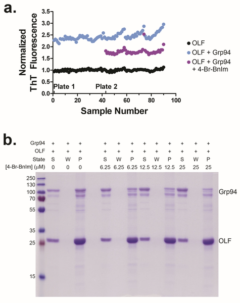
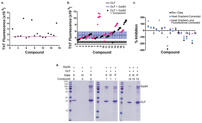
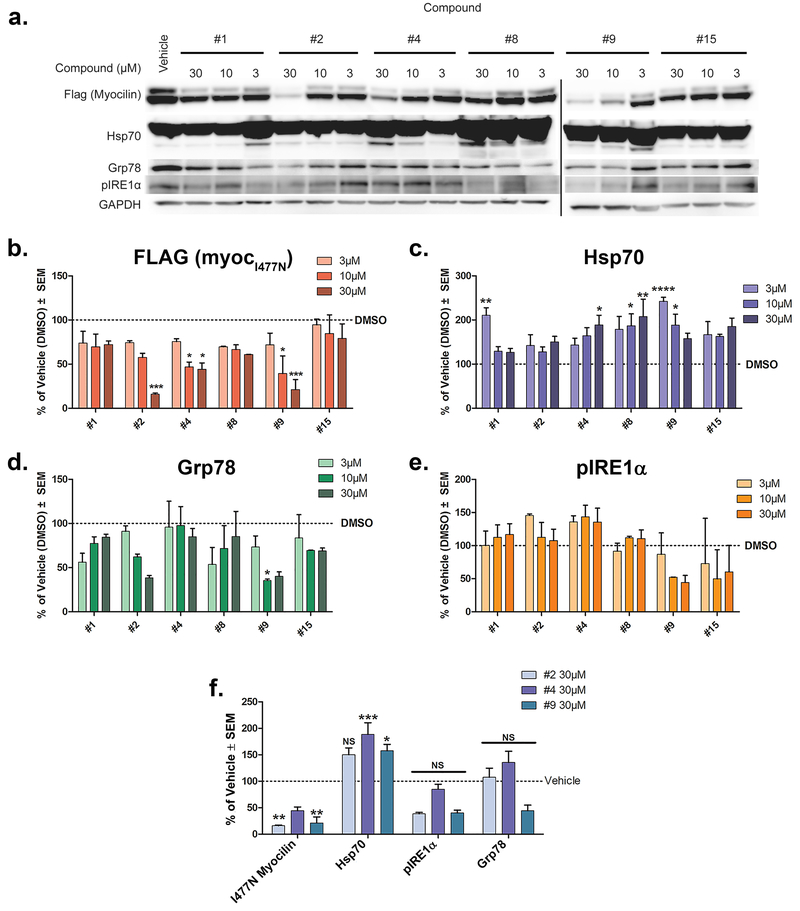
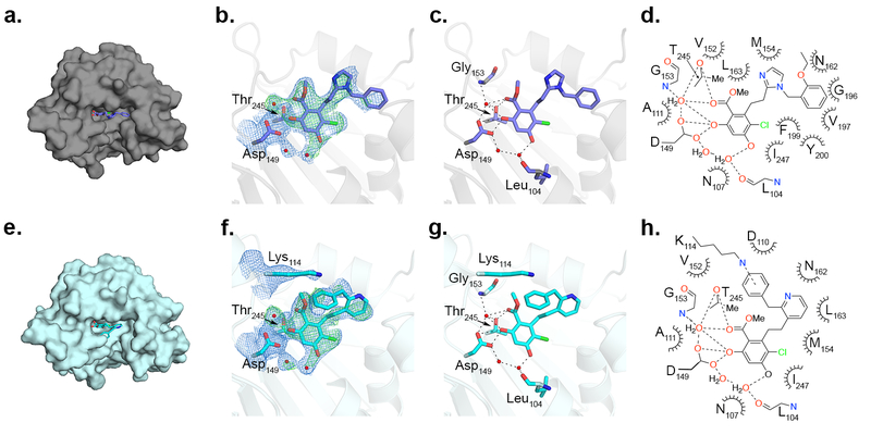
Gain-of-function mutations within the olfactomedin (OLF) domain of myocilin result in its toxic intracellular accumulation and hasten the onset of open-angle glaucoma. The absence of myocilin does not cause disease; therefore, strategies aimed at eliminating myocilin could lead to a successful glaucoma treatment. The endoplasmic reticulum Hsp90 paralog Grp94 accelerates OLF aggregation. Knockdown or pharmacological inhibition of Grp94 in cells facilitates clearance of mutant myocilin via a non-proteasomal pathway. Here, we expanded our support for targeting Grp94 over cytosolic paralogs Hsp90α and Hsp90β. We then developed a high-throughput screening assay to identify new chemical matter capable of disrupting the Grp94/OLF interaction. When applied to a blind, focused library of 17 Hsp90 inhibitors, our miniaturized single-read in vitro thioflavin T -based kinetics aggregation assay exclusively identified compounds that target the chaperone N-terminal nucleotide binding site. In follow up studies, one compound (2) decreased the extent of co-aggregation of Grp94 with OLF in a dose-dependent manner in vitro, and enabled clearance of the aggregation-prone full-length myocilin variant I477N in cells without inducing the heat shock response or causing cytotoxicity. Comparison of the co-crystal structure of compound 2 and another non-selective hit in complex with the N-terminal domain of Grp94 reveals a docking mode tailored to Grp94 and explains its selectivity. A new lead compound has been identified, supporting a targeted chemical biology assay approach to develop a protein degradation-based therapy for myocilin-associated glaucoma by selectively inhibiting Grp94.
Figures

Similar articles
-
Molecular Insights into Myocilin and Its Glaucoma-Causing Misfolded Olfactomedin Domain Variants.Acc Chem Res. 2021 May 4;54(9):2205-2215. doi: 10.1021/acs.accounts.1c00060. Epub 2021 Apr 13. Acc Chem Res. 2021. PMID: 33847483 Free PMC article. Review.
-
Different Grp94 components interact transiently with the myocilin olfactomedin domain in vitro to enhance or retard its amyloid aggregation.Sci Rep. 2019 Sep 4;9(1):12769. doi: 10.1038/s41598-019-48751-8. Sci Rep. 2019. PMID: 31484937 Free PMC article.
-
Exploiting the interaction between Grp94 and aggregated myocilin to treat glaucoma.Hum Mol Genet. 2014 Dec 15;23(24):6470-80. doi: 10.1093/hmg/ddu367. Epub 2014 Jul 15. Hum Mol Genet. 2014. PMID: 25027323 Free PMC article.
-
Glucose-regulated protein 94 triage of mutant myocilin through endoplasmic reticulum-associated degradation subverts a more efficient autophagic clearance mechanism.J Biol Chem. 2012 Nov 23;287(48):40661-9. doi: 10.1074/jbc.M112.384800. Epub 2012 Oct 3. J Biol Chem. 2012. PMID: 23035116 Free PMC article.
-
The effects of myocilin expression on functionally relevant trabecular meshwork genes: a mini-review.J Ocul Pharmacol Ther. 2014 Mar-Apr;30(2-3):202-12. doi: 10.1089/jop.2013.0218. Epub 2014 Feb 24. J Ocul Pharmacol Ther. 2014. PMID: 24564495 Free PMC article. Review.
Cited by
-
Amyloid fibrillation of the glaucoma associated myocilin protein is inhibited by epicatechin gallate (ECG).RSC Adv. 2022 Oct 14;12(45):29469-29481. doi: 10.1039/d2ra05061g. eCollection 2022 Oct 11. RSC Adv. 2022. PMID: 36320765 Free PMC article.
-
Glucose-regulated protein 94 (Grp94/gp96) in viral pathogenesis: Insights into its role and therapeutic potentials.Eur J Med Chem. 2025 Aug 5;292:117713. doi: 10.1016/j.ejmech.2025.117713. Epub 2025 Apr 30. Eur J Med Chem. 2025. PMID: 40319577 Free PMC article. Review.
-
Construction of miRNA-mRNA regulatory network indicates potential biomarkers for primary open-angle glaucoma.BMC Med Genomics. 2023 Nov 8;16(1):280. doi: 10.1186/s12920-023-01698-2. BMC Med Genomics. 2023. PMID: 37940950 Free PMC article.
-
Molecular Insights into Myocilin and Its Glaucoma-Causing Misfolded Olfactomedin Domain Variants.Acc Chem Res. 2021 May 4;54(9):2205-2215. doi: 10.1021/acs.accounts.1c00060. Epub 2021 Apr 13. Acc Chem Res. 2021. PMID: 33847483 Free PMC article. Review.
-
Physiological function of myocilin and its role in the pathogenesis of glaucoma in the trabecular meshwork (Review).Int J Mol Med. 2019 Feb;43(2):671-681. doi: 10.3892/ijmm.2018.3992. Epub 2018 Nov 20. Int J Mol Med. 2019. PMID: 30483726 Free PMC article. Review.
References
-
- Stone EM, Fingert JH, Alward WL, Nguyen TD, Polansky JR, Sunden SL, Nishimura D, Clark AF, Nystuen A, Nichols BE, Mackey DA, Ritch R, Kalenak JW, Craven ER, and Sheffield VC (1997) Identification of a gene that causes primary open angle glaucoma, Science 275, 668–670. - PubMed
-
- Hardy KM, Hoffman EA, Gonzalez P, McKay BS, and Stamer WD (2005) Extracellular trafficking of myocilin in human trabecular meshwork cells, J. Biol. Chem 280, 28917–28926. - PubMed
Publication types
MeSH terms
Substances
Grants and funding
LinkOut - more resources
Full Text Sources
Other Literature Sources
Medical
Miscellaneous
