. 2018 Mar;243(5):395-407.
doi: 10.1177/1535370218757458. Epub 2018 Feb 5.

Featured Article: Immunomodulatory effect of hemozoin on pneumocyte apoptosis via CARD9 pathway, a possibly retarding pulmonary resolution

Affiliations

Featured Article: Immunomodulatory effect of hemozoin on pneumocyte apoptosis via CARD9 pathway, a possibly retarding pulmonary resolution

Sitang Maknitikul et al. Exp Biol Med (Maywood). 2018 Mar.

Abstract

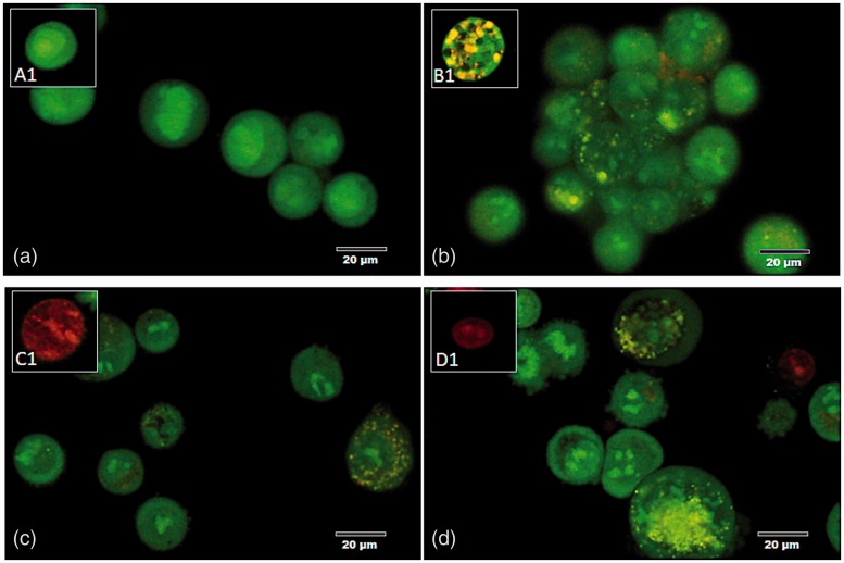
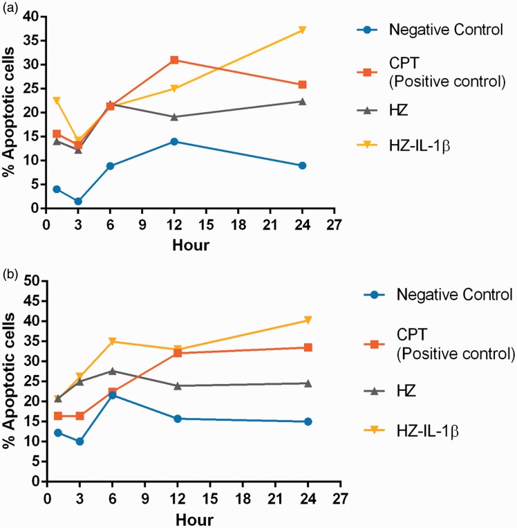
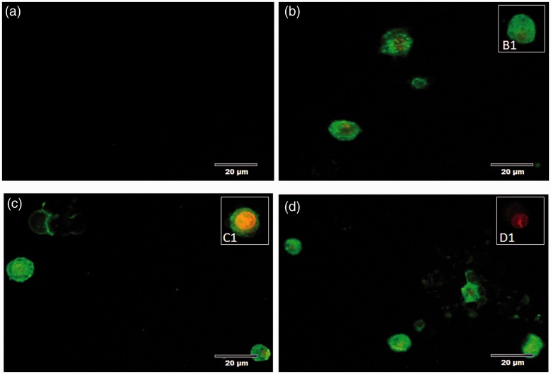
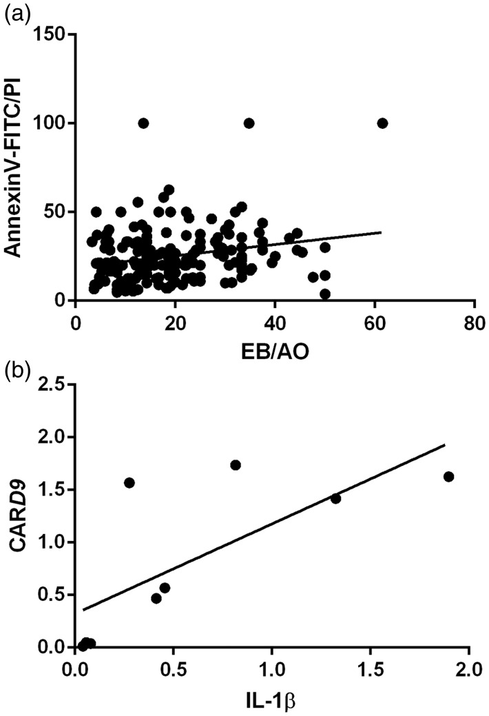
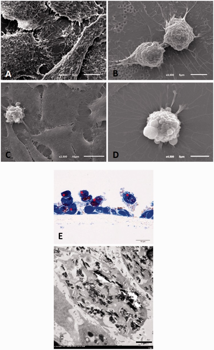
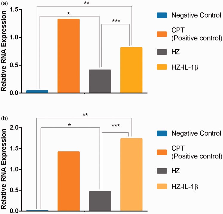
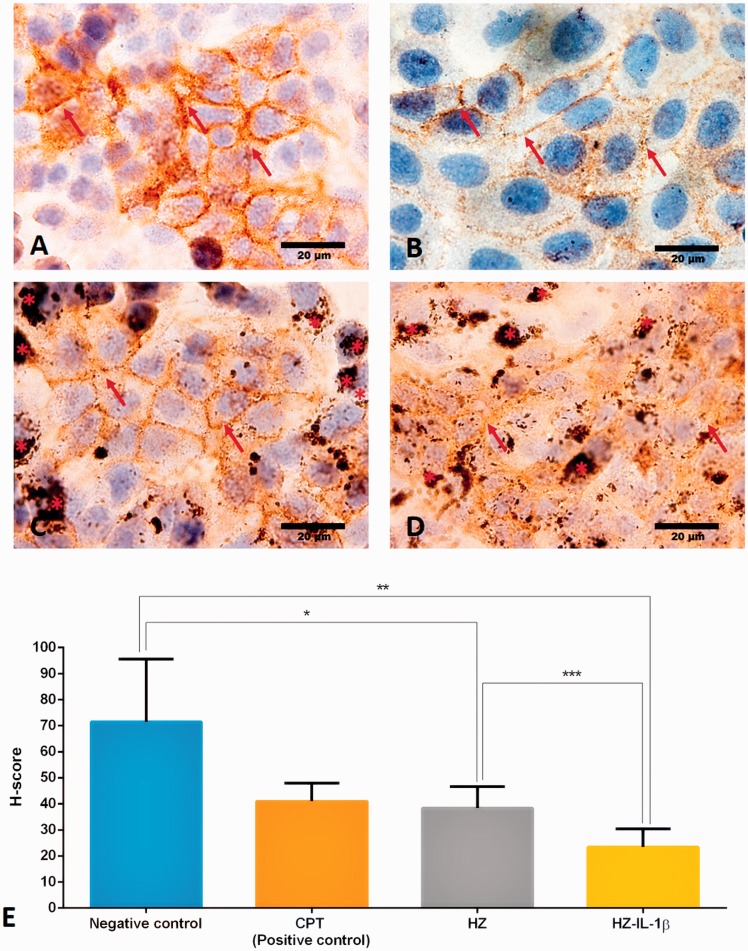
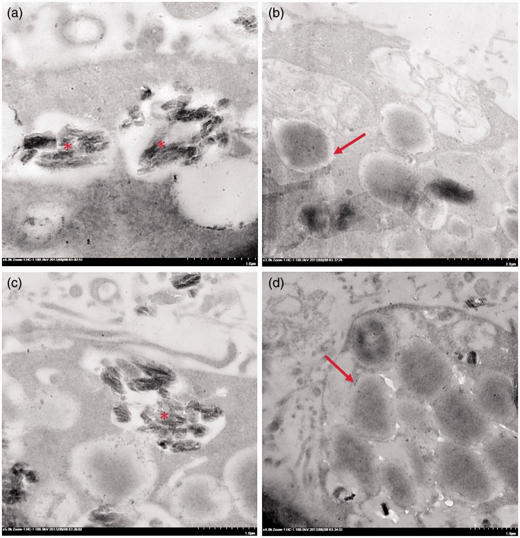
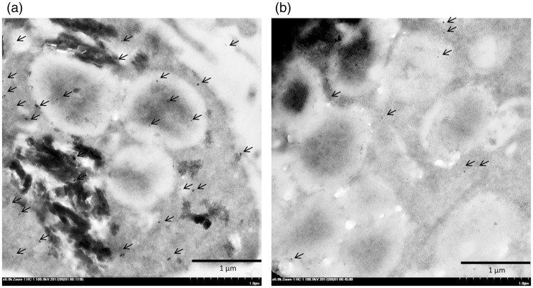
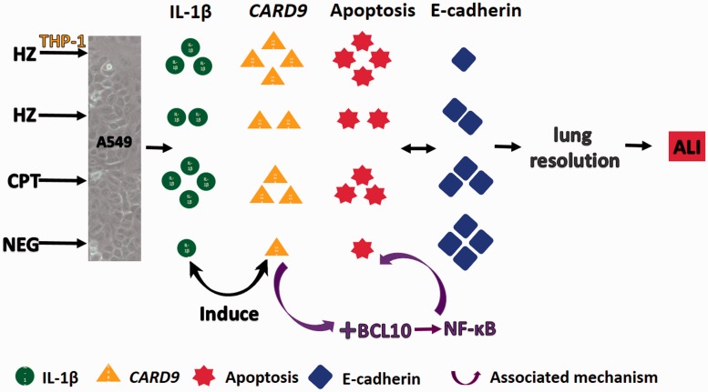
Plasmodium falciparum, the most virulent malaria parasite species, causes severe symptoms especially acute lung injury (ALI), of which characterized by alveolar epithelium and endothelium destruction and accelerated to blood-gas-barrier breakdown. Parasitized erythrocytes, endothelial cells, monocytes, and cytokines are all involved in this mechanism, but hemozoin (HZ), the parasitic waste from heme detoxification, also mainly contributes. In addition, it is not clear why type II pneumocyte proliferation, alveolar restorative stage, is rare in malaria-associated ALI. To address this, in vitro culture of A549 cells with Plasmodium HZ or with interleukin (IL)-1β triggered by HZ and monocytes (HZ-IL-1β) was conducted to determine their alveolar apoptotic effect using ethidium bromide/acridine orange staining, annexin-V-FITC/propidium iodide staining, and electron mircroscopic study. Caspase recruitment domain-containing protein 9 ( CARD9), the apoptotic regulator gene, and IL-1β were quantified by reverse-transcriptase PCR. Junctional cellular defects were characterized by immunohistochemical staining of E-cadherin. The results revealed that cellular apoptosis and CARD9 expression levels were extremely high 24 h after induction by HZ-IL-1β when compared to the HZ- and non-treated groups. E-cadherin was markedly down-regulated by HZ-IL-1β and HZ treatments. CARD9 expression was positively correlated with IL-1β expression and the number of apoptotic cells. Interestingly, the localization of HZ in the vesicular surfactant of apoptotic pneumocyte was also identified and submitted to be a cause of alveolar resolution abnormality. Thus, HZ triggers monocytes to produce IL-1β and induces pneumocyte type II apoptosis through CARD9 pathway in association with down-regulated E-cadherin, which probably impairs alveolar resolution in malaria-associated ALI. Impact statement The present work shows the physical and immunomodulatory properties of hemozoin on the induction of pneumocyte apoptosis in relation to IL-1β production through the CARD9 pathway. This occurrence may be a possible pathway for the retardation of lung resolution leading to blood-gas-barrier breakdown. Our findings lead to the understanding of the host-parasite relationship focusing on the dysfunction in ALI induced by HZ, a possible pathway of the recovering lung epithelial retardation in malaria-associated ARDS.

Keywords: Acute lung injury; CARD9; apoptosis; hemozoin; interleukin-1β.


Figures

Figure 1.
Figure 1.
Morphology of A549 apoptotic cells at five difference concentrations of HZ. The appropriate dose of HZ capable of inducing apoptosis in A549 cells was established by culturing the cells in the following concentrations of HZ: (a) 0 µM (negative control), (b) 2 µM, (c) 5 µM, (d) 10 µM, (e) 20 µM, and (f) 100 µM. Post induction, the intact cells showed large and cuboidal flattening and blue staining, whereas the apoptotic cells were small and starry like with hyperchromatic or dark nuclei. (A color version of this figure is available in the online journal.)
Figure 2.
Figure 2.
Morphology and EB/AO staining pattern of apoptotic cells. (a) A normal pneumocyte from the non-treated group with a green-stained appearance and intact nucleus. (b–d) The appearance of the apoptotic pneumocytes showed green to yellow staining with fragmented nuclei in the (b) CPT, (c) HZ, and (d) HZ-IL-1β treatment groups. (A color version of this figure is available in the online journal.)
Figure 3.
Figure 3.
Percentage of apoptotic pneumocytes treated with HZ. The number of apoptotic cells from EB/AO and annexin V-FITC/PI staining was calculated from line graphs. (a, b) At 1 h post-exposure, the group with the fastest cellular apoptotic induction was HZ-IL-1β. CPT and HZ alone groups were the next to reach high apoptosis levels after 6 and 12 h exposure times. At 24 h post-exposure, the HZ-IL-1β groups had higher numbers of apoptotic cells than those of the CPT-, HZ- and no-treatment groups. (A color version of this figure is available in the online journal.)
Figure 4.
Figure 4.
Morphology and annexin V-FITC/PI staining pattern of apoptotic cells. (a) In the no-treatment group, the intact pneumocytes were neither stained with annexin V-FITC nor PI, whereas the apoptotic pneumocytes were stained with annexin V-FITC with or without PI as shown in (b) CPT, (c) HZ, and (d) HZ with the THP-1 supernatant-treated group. (A color version of this figure is available in the online journal.)
Figure 5.
Figure 5.
Positive correlation between the apoptotic cell numbers determined by the different stains and IL-1β and CARD9 gene expression.
Figure 6.
Figure 6.
Fine morphological structure of A549 cells with or without apoptosis. The electron micrographs showed that (a) mature A549 cells were generally flat with irregular surfaces; (b) A549 progenitor cells were round with regular or irregular surfaces; (c, d) apoptotic A549 cells had blebs on their surfaces. (e) Semi-thin section of A549 cells with toluidine blue staining showing the HZ-treated apoptotic cells (*). HZ pigment deposited in the pneumocytes was frequently found in the HZ and HZ-IL-1β treatment groups (f). (A color version of this figure is available in the online journal.)
Figure 7.
Figure 7.
IL-1β and CARD9 mRNA expression in A549 cells by treatment group. HZ was able to up-regulate IL-1β and CARD9 levels in A549 cells. (a, b) HZ was able to induce apoptosis by its immunomodulatory activity but not in the non-treated group. CPT was the most effective treatment at inducing IL-1β genes (a), while HZ-IL-1β was the most effective treatment at inducing CARD9 gene expression (b). (A color version of this figure is available in the online journal.)
Figure 8.
Figure 8.
Immunohistochemical staining of E-cadherin in A549 cells by treatment group. A549 cells co-cultured with HZ or HZ-IL-1β were able to suppress E-cadherin expression. (a) Cells in the no-treatment group had intensive brown staining (arrow), whereas the groups treated with (b) CPT, (c) HZ, or (d) HZ-IL-1β rarely showed DAB staining accompanying HZ accumulation (*). (e) H-scores were calculated from the percentage of an area of expression/high power field and the intensity score. The results show that all the treatment groups experienced down-regulated E-cadherin expression. The HZ-IL-1β treatment group had the most pronounced suppression of E-cadherin expression in the pneumocytes, and the CPT and HZ treatment groups less so when compared with the no treatment group. (A color version of this figure is available in the online journal.)
Figure 9.
Figure 9.
Transmission electron micrograph of multivesicular bodies in pneumocytes exposed to HZ. Accumulation of HZ (*) showing lipid substitution in the multivesicular bodies (arrow).
Figure 10.
Figure 10.
Immunogold labeling of LC-3 in the surfactant vesicles. A number of LC-3 labeled gold particles (arrow) were found in the HZ-pigment-laden pneumocytes (a) as compared with the intact type II pneumocytes (b).
Figure 11.
Figure 11.
Possible mechanism for the retardation of lung resolution in HZ-induced ALI in relation to IL-1β and CARD9 regulation. (A color version of this figure is available in the online journal.)

References

    1. Wassmer SC, Grau GE. Severe malaria: what's new on the pathogenesis front? Int J Parasitol 2017; 47:145–52 - PMC - PubMed
    1. Sein KK, Maeno Y, Thuc HV, Anh TK, Aikawa M. Differential sequestration of parasitized erythrocytes in the cerebrum and cerebellum in human cerebral malaria. Am J Trop Med Hyg 1993; 48:504–11 - PubMed
    1. Nguansangiam S, Day NP, Hien TT, Mai NT, Chaisri U, Riganti M, Dondorp AM, Lee SJ, Phu NH, Turner GD, White NJ, Ferguson DJ, Pongponratn E. A quantitative ultrastructural study of renal pathology in fatal Plasmodium falciparum malaria. Trop Med Int Health 2007; 12:1037–50 - PubMed
    1. Taylor WR, Hanson J, Turner GD, White NJ, Dondorp AM. Respiratory manifestations of malaria. Chest 2012; 142:492–505 - PubMed
    1. Mohan A, Sharma SK, Bollineni S. Acute lung injury and acute respiratory distress syndrome in malaria. J Vector Borne Dis 2008; 45:179–93 - PubMed

Publication types

MeSH terms