. 2018 Feb 5;17(1):17.
doi: 10.1186/s12934-018-0868-0.

Establishment of a yeast-based VLP platform for antigen presentation

Affiliations

Establishment of a yeast-based VLP platform for antigen presentation

David Wetzel et al. Microb Cell Fact. .

Abstract

Background: Chimeric virus-like particles (VLP) allow the display of foreign antigens on their surface and have proved valuable in the development of safe subunit vaccines or drug delivery. However, finding an inexpensive production system and a VLP scaffold that allows stable incorporation of diverse, large foreign antigens are major challenges in this field.

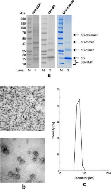
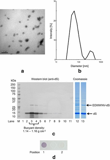
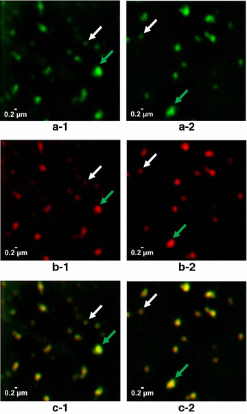
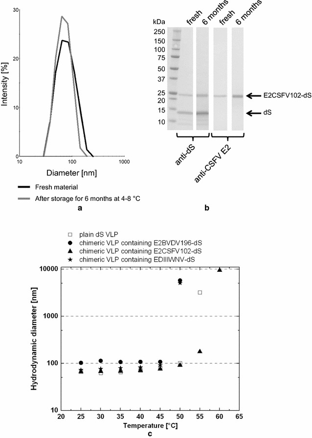
Results: In this study, a versatile and cost-effective platform for chimeric VLP development was established. The membrane integral small surface protein (dS) of the duck hepatitis B virus was chosen as VLP scaffold and the industrially applied and safe yeast Hansenula polymorpha (syn. Pichia angusta, Ogataea polymorpha) as the heterologous expression host. Eight different, large molecular weight antigens of up to 412 amino acids derived from four animal-infecting viruses were genetically fused to the dS and recombinant production strains were isolated. In all cases, the fusion protein was well expressed and upon co-production with dS, chimeric VLP containing both proteins could be generated. Purification was accomplished by a downstream process adapted from the production of a recombinant hepatitis B VLP vaccine. Chimeric VLP were up to 95% pure on protein level and contained up to 33% fusion protein. Immunological data supported surface exposure of the foreign antigens on the native VLP. Approximately 40 mg of chimeric VLP per 100 g dry cell weight could be isolated. This is highly comparable to values reported for the optimized production of human hepatitis B VLP. Purified chimeric VLP were shown to be essentially stable for 6 months at 4 °C.

Conclusions: The dS-based VLP scaffold tolerates the incorporation of a variety of large molecular weight foreign protein sequences. It is applicable for the display of highly immunogenic antigens originating from a variety of pathogens. The yeast-based production system allows cost-effective production that is not limited to small-scale fundamental research. Thus, the dS-based VLP platform is highly efficient for antigen presentation and should be considered in the development of future vaccines.

Keywords: Animal infectious diseases; Antibiotic-free; Antigen presentation; Chimeric virus-like particles; DHBV; Hansenula polymorpha; Pichia angusta; Virus-like particles.


Figures

Fig. 1
Fig. 1
Map of the novel expression plasmid pB14-2xFPMT-dS carrying two expression cassettes specifically tailored to heterologous co-production of the dS and a fusion protein for chimeric VLP production with H. polymorpha. The dS encoding gene is stably inserted. The ORF encoding the desired fusion protein is to be inserted using EcoRI and BamHI sites. The S. cerevisiae URA3 gene was used for selection of bacteria and yeast. Further features: ori, origin of replication; HARS1, H. polymorpha autonomously replicating sequence 1; FMD-P, FMD promoter; MOX, MOX terminator both derived from H. polymorpha genome
Fig. 2
Fig. 2
Western blot analysis of crude cell lysates probed with anti-dS mAB 7C12. Samples originated from 17 different recombinant H. polymorpha strains co-producing the dS and the fusion protein E2CSFV102-dS (lanes 1, 3–17) or producing only the dS (lane 2, strain A#299). Indicated strain generation strategies (I), (II) and (III) refer to the methods described in the text. Strains A#299 and D#79 (highlighted by arrows) were used for VLP production. M, molecular weight marker
Fig. 3
Fig. 3
Protein deglycosylation assay of crude cell lysates derived from recombinant H. polymorpha strains D#53 (a) and D#73 (b) co-producing the dS and the fusion proteins E2CSFV337-dS or E2CSFV184-dS, respectively. Samples were analyzed by Western blot probed with anti-dS mAB 7C12 with (lanes 1a and 1b) or without (lanes 2a and 2b) previous EndoH treatment. M, molecular weight marker
Fig. 4
Fig. 4
Western blot analysis of crude cell lysates probed with anti-CSFV E2 mAB. Samples originated from strains D#53 (lane 1), D#73 (lane 2) and D#79 (lane 3) co-producing the dS and the indicated fusion protein and A#299 (lane 4) producing the dS but no fusion protein. M, molecular weight marker
Fig. 5
Fig. 5
CsCl density gradient separation of HCP from plain dS VLP derived from recombinant H. polymorpha strain A#299 analyzed by Western blot. The ultracentrifugation tube was divided into 11 fractions with increasing density from fraction 1–11. Top: unspecific staining of proteins (Ponceau S). Bottom: anti-dS immunostaining using 7C12 mAB. M, molecular weight marker
Fig. 6
Fig. 6
Analysis of plain dS VLP purified from recombinant H. polymorpha strain A#299. a Western blot probed with anti-HCP serum (10 µg protein loaded) or anti-dS mAB 7C12 (1 µg protein loaded), respectively and Coomassie stained PAA gel analysis (10 µg protein loaded); M, molecular weight marker; HMF, higher mobility forms. b TEM images at 50,000-fold (top) and 250,000-fold (bottom) magnification. c DLS data after regularization analysis
Fig. 7
Fig. 7
Characterization of chimeric VLP isolated from strain T#3-3 co-producing dS and EDIIIWNV-dS and desalted by SEC. a TEM image after negative staining (100,000-fold magnification); b DLS data after regularization analysis; c lanes 1–11: Western blot analysis of fractions harvested from analytical CsCl density gradient separation (density increases gradually from lanes 1–11) probed with anti-dS mAB 7C12; Coomassie stained PAA gels for analysis of final VLP preparations after desalting by dialysis (lane 12) or SEC (lane 13), 10 µg protein loaded; d dot blot analysis of the native sample desalted by SEC and probed with anti-WNV mAB, position 1: chimeric VLP displaying the WNV antigen, position 2: plain dS VLP as a negative control
Fig. 8
Fig. 8
Evaluation of chimeric VLP formation from material originated from strain D#79 co-producing dS and E2CSFV102-dS. a Lanes 1–11: Western blot analysis of fractions harvested from CsCl density gradient separation (density increases gradually from lanes 1–11), probed with anti-dS mAB 7C12; Lane 12: Coomassie stained PAA gel of pooled and desalted fractions 3 and 4. b TEM image after negative staining (100,000-fold magnification); c DLS data after regularization analysis
Fig. 9
Fig. 9
Chimeric VLP isolated from strain D#79 composed of dS and E2CSFV102-dS were analyzed under native conditions by N-SIM. Two series of images obtained from the same sample are presented showing fluorescence immunolabeling of dS in green (a-1, a-2), CSFV E2 antigen in red (b-1, b-2) and co-localization of the two labels in superimposed images in yellow (c-1, c-2). In each series of images two spots were consistently marked by arrows: signals of the size expected for individual VLP (white); largest signals in the respective frame (green)
Fig. 10
Fig. 10
Stability assessment of chimeric VLP. a, b DLS and Western blot analysis of real time stability experiment of purified VLP composed of dS and E2CSFV102-dS, isolated from strain D#79 and formulated at mg mL−1 protein concentration in desalting buffer. Data of fresh sample analysis are compared to data collected after 6 months of storage at 4–8 °C by independent but volume-normalized Western blot analysis. c DLS analysis of plain dS VLP and different chimeric VLP analyzed during step-wise increasing temperature allowing 5 min equilibration time in between the measurements

References

    1. McAleer WJ, Buynak EB, Maigetter RZ, Wampler DE, Miller WJ, Hilleman MR. Human hepatitis B vaccine from recombinant yeast. Nature. 1984;307:178–180. doi: 10.1038/307178a0. - DOI - PubMed
    1. Zhao Q, Li S, Yu H, Xia N, Modis Y. Virus-like particle-based human vaccines: quality assessment based on structural and functional properties. Trends Biotechnol. 2013;11:654–663. doi: 10.1016/j.tibtech.2013.09.002. - DOI - PubMed
    1. Rohovie MJ, Nagasawa M, Swartz JR. Virus-like particles: next-generation nanoparticles for targeted therapeutic delivery. Bioeng Transl Med. 2016;2:43–57. - PMC - PubMed
    1. Yildiz I, Shukla S, Steinmetz NF. Applications of viral nanoparticles in medicine. Curr Opin Biotechnol. 2011;22:901–908. doi: 10.1016/j.copbio.2011.04.020. - DOI - PMC - PubMed
    1. Liu F, Ge S, Li L, Wi X, Liu Z, Wang Z. Virus-like particles: potential veterinary vaccine immunogens. Res Vet Sci. 2012;93:553–559. doi: 10.1016/j.rvsc.2011.10.018. - DOI - PubMed

MeSH terms

Substances