. 2018 Feb 5;11(1):12.
doi: 10.1186/s13048-018-0385-0.

Sialylation of EGFR by the ST6Gal-I sialyltransferase promotes EGFR activation and resistance to gefitinib-mediated cell death

Affiliations

Sialylation of EGFR by the ST6Gal-I sialyltransferase promotes EGFR activation and resistance to gefitinib-mediated cell death

Colleen M Britain et al. J Ovarian Res. .

Abstract

Background: The ST6Gal-I sialyltransferase is upregulated in numerous cancers, and high expression of this enzyme correlates with poor patient prognosis in various malignancies, including ovarian cancer. Through its sialylation of a select cohort of cell surface receptors, ST6Gal-I modulates cell signaling to promote tumor cell survival. The goal of the present study was to investigate the influence of ST6Gal-I on another important receptor that controls cancer cell behavior, EGFR. Additionally, the effect of ST6Gal-I on cancer cells treated with the common EGFR inhibitor, gefitinib, was evaluated.

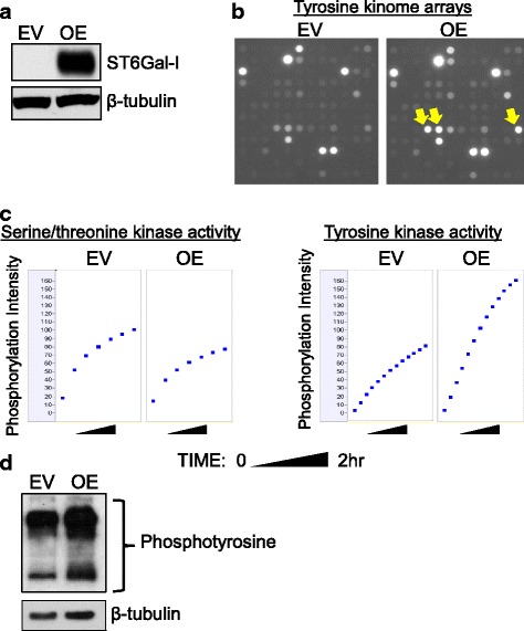
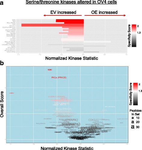
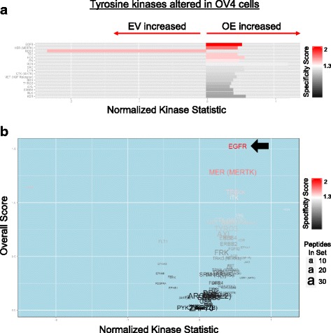
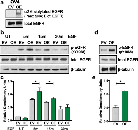
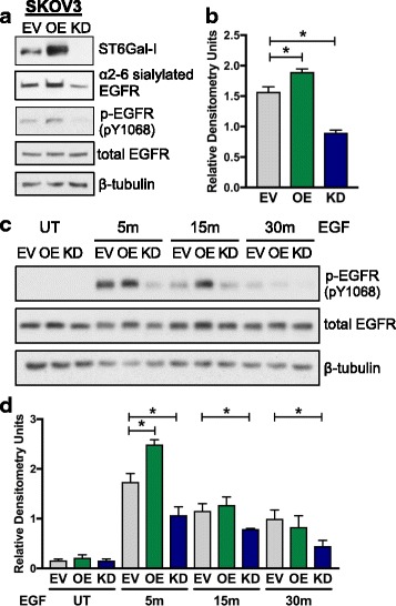
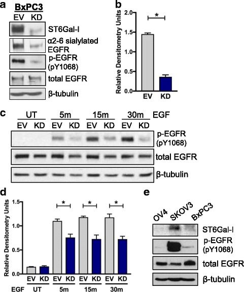
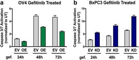
Results: Using the OV4 ovarian cancer cell line, which lacks endogenous ST6Gal-I expression, a kinomics assay revealed that cells with forced overexpression of ST6Gal-I exhibited increased global tyrosine kinase activity, a finding confirmed by immunoblotting whole cell lysates with an anti-phosphotyrosine antibody. Interestingly, the kinomics assay suggested that one of the most highly activated tyrosine kinases in ST6Gal-I-overexpressing OV4 cells was EGFR. Based on these findings, additional analyses were performed to investigate the effect of ST6Gal-I on EGFR activation. To this end, we utilized, in addition to OV4 cells, the SKOV3 ovarian cancer cell line, engineered with both ST6Gal-I overexpression and knockdown, as well as the BxPC3 pancreatic cancer cell line with knockdown of ST6Gal-I. In all three cell lines, we determined that EGFR is a substrate of ST6Gal-I, and that the sialylation status of EGFR directly correlates with ST6Gal-I expression. Cells with differential ST6Gal-I expression were subsequently evaluated for EGFR tyrosine phosphorylation. Cells with high ST6Gal-I expression were found to have elevated levels of basal and EGF-induced EGFR activation. Conversely, knockdown of ST6Gal-I greatly attenuated EGFR activation, both basally and post EGF treatment. Finally, to illustrate the functional importance of ST6Gal-I in regulating EGFR-dependent survival, cells were treated with gefitinib, an EGFR inhibitor widely used for cancer therapy. These studies showed that ST6Gal-I promotes resistance to gefitinib-mediated apoptosis, as measured by caspase activity assays.

Conclusion: Results herein indicate that ST6Gal-I promotes EGFR activation and protects against gefitinib-mediated cell death. Establishing the tumor-associated ST6Gal-I sialyltransferase as a regulator of EGFR provides novel insight into the role of glycosylation in growth factor signaling and chemoresistance.

Keywords: Epidermal growth factor receptor (EGFR) cell signaling; Gefitinib; Glycosylation; Kinomics; Tumor cell biology; Tyrosine kinase; β-galactoside α2-6 sialyltransferase 1 (ST6GAL1).


Conflict of interest statement

Ethics approval and consent to participate

Not applicable.

Consent for publication

Not applicable.

Competing interests

The authors declare that they have no competing interests.

Publisher’s Note

Springer Nature remains neutral with regard to jurisdictional claims in published maps and institutional affiliations.

Figures

Fig. 1
Fig. 1
ST6Gal-I promotes an increase in overall tyrosine kinase activity. a. OV4 cells were stably transduced with lentivirus encoding ST6Gal-I, and ST6Gal-I overexpression (OE) was confirmed by immunoblotting (EV = empty vector control). b. Whole array image capture at final prewash cycle number 92 of PTK array illustrating changes in tyrosine phosphorylation with qualitatively selected altered spots (yellow arrows) increased in ST6Gal-I OV4 OE cells as compared to EV cells. c. Whole chip comparative OE and EV array-mean phosphorylation intensity (y axis per cell) over time in the kinetic/prewash cycles (x axis per cell) for both the STK (left 2 panels) and PTK (right two panels) arrays. d. EV or OE cells were immunoblotted with an anti-phosphotyrosine antibody
Fig. 2
Fig. 2
ST6Gal-I overexpression attenuates serine/threonine kinase activity. Bar and volcano plots of kinases with altered STK activity in OV4 OE vs. EV cells. a. Bar plot of serine/threonine kinases identified with the BioNavigator UpKin STK PamApp v14.0 (PamGene) scored as increased (rightward) or decreased (leftward) in OE relative to EV. Length of bar indicates extent of change (KSTAT; Kinase Statistic) and color of bar indicates specificity of each kinase to the predicted phosphosites used to measure its respective activity. b. In the volcano plot, kinases are similarly scored as increased or decreased (y axis) in OE relative to EV, colored by specificity. A combined KSTAT + specificity score (x axis) is denoted, with text size indicating the number of seed peptides used to identify that kinase
Fig. 3
Fig. 3
Tyrosine kinases, including EGFR, are more activated in cells with ST6Gal-I overexpression. Bar and volcano plots of kinases with altered PTK activity in OV4 OE vs. EV cells. a. Bar plot of tyrosine kinases identified with the BioNavigator UpKin PTK PamApp v8.0 (PamGene) scored as increased (rightward) or decreased (leftward) in OE relative to EV. Length of bar indicates extent of change (KSTAT; Kinase Statistic) and color of bar indicates specificity of each kinase to the predicted phosphosites used to measure its respective activity. b. In the volcano plot, kinases are similarly scored as increased or decreased (y axis) in OE relative to EV, colored by specificity. A combined KSTAT + specificity score (x axis) is denoted, with text size indicating the number of seed peptides used to identify that kinase
Fig. 4
Fig. 4
EGFR phosphorylation is enhanced in OV4 ovarian cancer cells with ST6Gal-I overexpression. a. To measure levels of α2-6 sialylated EGFR, cell lysates were incubated with SNA-agarose. SNA is a lectin that specifically recognizes α2-6 sialic acids. Sialylated proteins were precipitated and then immunoblotted for EGFR. b. Representative p-EGFR immunoblot of EV or OE cells treated with EGF for 5, 15 and 30 min. c. Densitometric analysis of three independent blots of p-EGFR in EGF-treated OV4 cells, with values normalized to the β-tubulin loading control. d. Representative p-EGFR immunoblot of EV or OE cells to evaluate basal EGFR phosphorylation. e. Densitometric analysis of three independent blots of basal p-EGFR in OV4 cells. *, p < 0.05
Fig. 5
Fig. 5
EGFR phosphorylation is enhanced in SKOV3 ovarian cancer cells with ST6Gal-I overexpression, but decreased in cells with ST6Gal-I knockdown. a. SKOV3 cells were stably transduced with lentivirus encoding human ST6Gal-I, or alternatively, shRNA for ST6Gal-I. ST6Gal-I overexpression (OE) and knockdown (KD) were confirmed by immunoblotting. EV, OE or KD cell lysates were precipitated by SNA-agarose and immunoblotted for EGFR to detect levels of α2-6 sialylated EGFR. Total cell lysates were immunoblotted for activated EGFR (p-EGFR, pY1068) or total EGFR. b. Densitometric analysis of three independent blots of basal p-EGFR in SKOV3 cells (normalized to β-tubulin). c. Representative p-EGFR immunoblot of EV, OE or KD cells treated with EGF for 5, 15 and 30 min. d. Densitometric analysis of three independent blots of p-EGFR in EGF-treated SKOV3 cells. *, p < 0.05
Fig. 6
Fig. 6
Knockdown of ST6Gal-I diminishes EGFR phosphorylation in BxPC3 pancreatic cancer cells. a. Using lentivirus, BxPC3 cells were stably transduced with shRNA for ST6Gal-I, and ST6Gal-I knockdown (KD) was confirmed by immunoblotting. EV or KD cell lysates were precipitated by SNA agarose and immunoblotted for EGFR to detect α2-6 sialylated EGFR. Total cell lysates were immunoblotted for activated EGFR (p-EGFR, pY1068) or total EGFR. b. Densitometric analysis of three independent blots of basal p-EGFR in BxPC3 cells (normalized to β-tubulin). c. Representative p-EGFR immunoblot of EV or KD cells treated with EGF for 5, 15 and 30 min. d. Densitometric analysis of three independent blots of p-EGFR in EGF-treated BxPC3 cells. e. Total cell lysates from EV cells of the OV4, SKOV3 and BxPC3 lines were immunoblotted for ST6Gal-I, p-EGFR (Y1068) or total EGFR. *, p < 0.05
Fig. 7
Fig. 7
ST6Gal-I activity protects cancer cells against gefitinib-mediated apoptosis. a. OV4 EV or OE cells and b. BxPC3 EV or KD cells were treated with gefitinib for 24, 48 and 72 h and analyzed for apoptosis via a luminescence assay that detects caspase 3 and 7 activity. Caspase activity in geftinib-treated cells was normalized to caspase activity in untreated cells. Data shown are from a representative experiment, with at least 2 independent experiments performed for each cell line. Graph depicts means ± S.D

References

    1. Dall’Olio F, Chiricolo M. Sialyltransferases in cancer. Glycoconj J. 2001;18:841–850. doi: 10.1023/A:1022288022969. - DOI - PubMed
    1. Varki NM, Varki A. Diversity in cell surface sialic acid presentations: implications for biology and disease. Laboratory investigation; a journal of technical methods and pathology. Lab Investig. 2007;87(9):851–857. doi: 10.1038/labinvest.3700656. - DOI - PMC - PubMed
    1. Schultz MJ, Swindall AF, Bellis SL. Regulation of the metastatic cell phenotype by sialylated glycans. Cancer Metastasis Rev. 2012;31(3-4):501–518. doi: 10.1007/s10555-012-9359-7. - DOI - PMC - PubMed
    1. Swindall AF, Londono-Joshi AI, Schultz MJ, Fineberg N, Buchsbaum DJ, Bellis SL. ST6Gal-I protein expression is upregulated in human epithelial tumors and correlates with stem cell markers in normal tissues and colon cancer cell lines. Cancer Res. 2013;73(7):2368–2378. doi: 10.1158/0008-5472.CAN-12-3424. - DOI - PMC - PubMed
    1. Schultz MJ, Holdbrooks AT, Chakraborty A, Grizzle WE, Landen CN, Buchsbaum DJ, et al. The tumor-associated glycosyltransferase ST6Gal-I regulates stem cell transcription factors and confers a cancer stem cell phenotype. Cancer Res. 2016;76(13):3978–3988. doi: 10.1158/0008-5472.CAN-15-2834. - DOI - PMC - PubMed

MeSH terms