. 2018 Feb 5;9(1):30.
doi: 10.1186/s13287-018-0788-2.

Embryonic stem cell-derived cardiomyocytes for the treatment of doxorubicin-induced cardiomyopathy

Affiliations

Embryonic stem cell-derived cardiomyocytes for the treatment of doxorubicin-induced cardiomyopathy

Danúbia Silva Dos Santos et al. Stem Cell Res Ther. .

Abstract

Background: Doxorubicin (Dox) is a chemotherapy drug with limited application due to cardiotoxicity that may progress to heart failure. This study aims to evaluate the role of cardiomyocytes derived from mouse embryonic stem cells (CM-mESCs) in the treatment of Dox-induced cardiomyopathy (DIC) in mice.

Methods: The mouse embryonic stem cell (mESC) line E14TG2A was characterized by karyotype analysis, gene expression using RT-PCR and immunofluorescence. Cells were transduced with luciferase 2 and submitted to cardiac differentiation. Total conditioned medium (TCM) from the CM-mESCs was collected for proteomic analysis. To establish DIC in CD1 mice, Dox (7.5 mg/kg) was administered once a week for 3 weeks, resulting in a cumulative Dox dose of 22.5 mg/kg. At the fourth week, a group of animals was injected intramyocardially with CM-mESCs (8 × 10⁵ cells). Cells were tracked by a bioluminescence assay, and the body weight, echocardiogram, electrocardiogram and number of apoptotic cardiomyocytes were evaluated.

Results: mESCs exhibited a normal karyotype and expressed pluripotent markers. Proteomic analysis of TCM showed proteins related to the negative regulation of cell death. CM-mESCs presented ventricular action potential characteristics. Mice that received Dox developed heart failure and showed significant differences in body weight, ejection fraction (EF), end-systolic volume (ESV), stroke volume (SV), heart rate and QT and corrected QT (QTc) intervals when compared to the control group. After cell or placebo injection, the Dox + CM-mESC group showed significant increases in EF and SV when compared to the Dox + placebo group. Reduction in ESV and QT and QTc intervals in Dox + CM-mESC-treated mice was observed at 5 or 30 days after cell treatment. Cells were detected up to 11 days after injection. The Dox + CM-mESC group showed a significant reduction in the percentage of apoptotic cardiomyocytes in the hearts of mice when compared to the Dox + placebo group.

Conclusions: CM-mESC transplantation improves cardiac function in mice with DIC.

Keywords: Cardiomyocytes derived from mouse embryonic stem cells; Cell therapy; Dox-induced cardiomyopathy; Heart failure.


Conflict of interest statement

Ethics approval and consent to participate

This study was approved by the Ethics Committee for Animal Use of the Federal University of Rio de Janeiro under number IBCCF 171/13.

Consent for publication

Not applicable.

Competing interests

The authors declare that they have no competing interests.

Publisher’s Note

Springer Nature remains neutral with regard to jurisdictional claims in published maps and institutional affiliations.

Figures
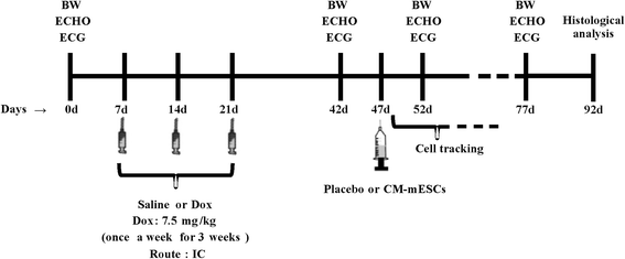
Fig. 1
Fig. 1
Experimental design. Schematic timeline of the study design. BW body weight, ECHO echocardiogram, ECG electrocardiogram, Dox doxorubicin, IC intracavitary, CM-mESC cardiomyocytes derived from mouse embryonic stem cell
Fig. 2
Fig. 2
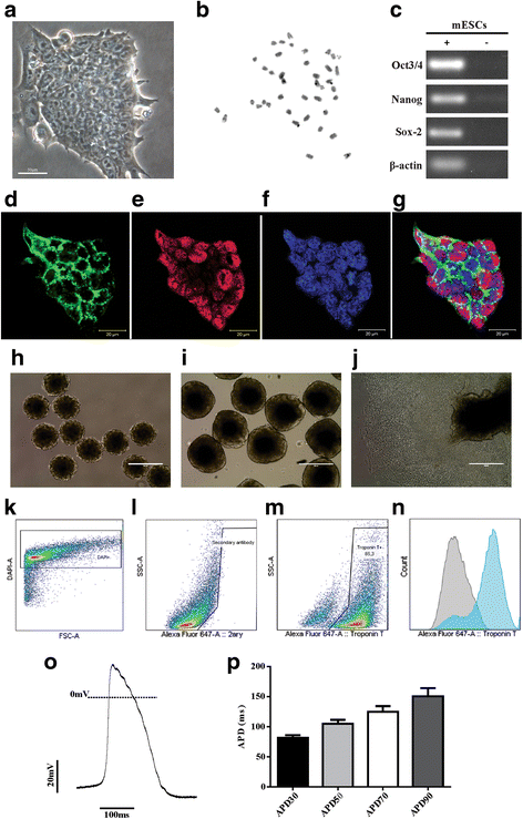
Characterization of undifferentiated mESC line E14TG2A, embryoid body formation and cardiac differentiation. a mESCs in culture. b Karyotype analysis showing 40 chromosomes. c Expression of undifferentiated mESC transcription factors by RT-PCR: Oct3/4, Nanog and Sox-2. dg Expression of pluripotency markers in mESCs by immunofluorescence: cells positive for d SSEA-1 (green) and e Oct3/4 (red); f nuclei labeled with Topro (blue); and g merged image. h, i Embryoid bodies (EBs) in suspension after 2 and 5 days of differentiation. j Adhered EBs after 8 days of differentiation with beating cells. kn Flow cytometry analysis on day 14: representative graphs of k DAPI-positive differentiated cells used to isolate permeabilized cells only, l differentiated cells stained only with the secondary antibody and m cardiac troponin T-positive differentiated cells; and n histogram overlay of differentiated cells stained with secondary antibody (gray) and cells expressing cardiac troponin t (blue). o Representative trace of ventricular action potential. p Action potential duration at 30, 50, 70 and 90% of repolarization (APD30, APD50, APD70 and APD90, respectively; n = 8). Scale bars: a 50 μm, dg 20 μm. mESC mouse embryonic stem cell, DAPI 4′,6-diamidino-2-phenylindole, FSC forward scatter, SSC side scatter
Fig. 3
Fig. 3
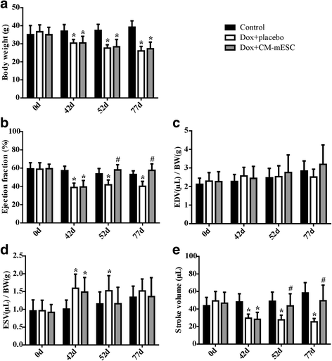
Assessment of cardiac function in DIC mice transplanted with CM-mESCs or placebo. Measures performed before (corresponding to day 0) and after (corresponding to day 42) Dox or saline administration, and after cell or placebo injection (corresponding to days 52 and 77, respectively). a Analysis of body weight (BW), b ejection fraction (EF), c, d end-diastolic volume (EDV) and end-systolic volume (ESV) corrected by BW, and e stroke volume (SV). Two-way ANOVA with Bonferroni post test: *p < 0.05 compared to the control group; #p < 0.05 compared to the Dox + placebo group. Data shown as mean ± standard deviation; n = 18 for the control group, n = 13 for the Dox + placebo group and n = 10 for the Dox + CM-mESC group. Dox doxorubicin, CM-mESC cardiomyocytes derived from mouse embryonic stem cell, days
Fig. 4
Fig. 4
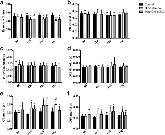
Assessment of electrical activity in DIC mice transplanted with CM-mESCs or placebo. Measures performed before (corresponding to day 0) and after (corresponding to day 42) Dox or saline administration, and after cell or placebo injection (corresponding to days 52 and 77, respectively). a Analysis of heart rate (HR), b PR interval, c P-wave duration, d QRS duration, e QT interval, and f QTc interval. Two-way ANOVA with a Bonferroni post test: *p < 0.05 compared to the control group; #p < 0.05 compared to the Dox + placebo group. Data shown as mean ± standard deviation; n = 18 for the control group, n = 13 for the Dox + placebo group and n = 10 for the Dox + CM-mESC group. Dox doxorubicin, CM-mESC cardiomyocytes derived from mouse embryonic stem cell, d days, QTc corrected QT
Fig. 5
Fig. 5
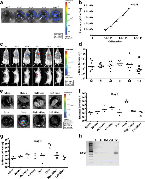
Cell tracking by bioluminescence. a Evaluation of luminescent signal intensity in CM-mESCs transduced with the luciferase 2 gene in vitro. Numbers shown above the wells indicate numbers of cells. b Signal increased with increasing cell number in a linear fashion (r2 = 0.98). c Representative images of CM-mESCs transduced with luciferase 2 after intramyocardial injection in DIC mice. Signal located in a region anatomically compatible with the heart in mice injected with CM-mESCs for up to 11 days after injection. d Quantification of luminescence in radiance units shows a decrease in the signal as time progresses. e Ex-vivo images and f, g quantification (n = 3 per day) of the luminescent signal demonstrating that cells remained in the heart at 1 day and 4 days after implantation, respectively. Signal lower in other organs (spleen, bladder, right lung, left lung, liver, right kidney and left kidney). h Presence of the luciferase 2 gene by PCR at 1, 4 and 11 days after cell injection. Luciferase 2 not observed at 45 days after injection. d days, PC positive control
Fig. 6
Fig. 6
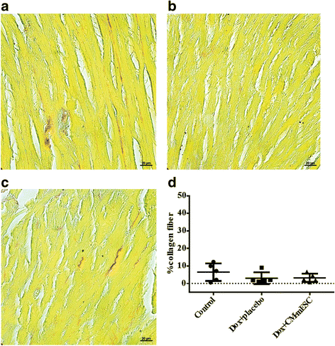
Histological analysis of the heart. ac Representative images of histological sections stained with Sirius Red: a control group, b Dox + placebo group and c Dox + CM-mESC group after 45 days of cell therapy. d No changes in myocardial collagen fiber content. One-way ANOVA with a Bonferroni post test. Data shown as mean ± standard deviation; n = 5 for each group. Dox doxorubicin, CM-mESC cardiomyocytes derived from mouse embryonic stem cell
Fig. 7
Fig. 7
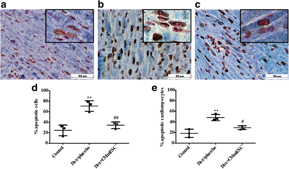
Apoptosis quantification in the heart. ac Representative images of tissue sections stained with TUNEL (brown), cardiac troponin T (blue) and Safranin O (red) (nonapoptotic nuclei) at 45 days after cell therapy: a control group, b Dox + placebo group and c Dox + CM-mESC group. Quantification of d total apoptotic cells and e apoptotic cardiomyocytes. One-way ANOVA with a Bonferroni post test: **p < 0.01 compared to the control group; #p < 0.05, ##p < 0.01 compared to the Dox + placebo group. Data shown as mean ± standard deviation; n = 3 for each group. Dox doxorubicin, CM-mESC cardiomyocytes derived from mouse embryonic stem cell

Similar articles

Cited by

References

    1. Minotti G, Menna P, Salvatorelli E, et al. Anthracyclines: molecular advances and pharmacologic developments in antitumor activity and cardiotoxicity. Pharmacol Rev. 2004;56:185–229. doi: 10.1124/pr.56.2.6. - DOI - PubMed
    1. Takemura G, Fujiwara H. Doxorubicin-induced cardiomyopathy from the cardiotoxic mechanisms to management. Prog Cardiovasc Dis. 2007;49(5):330–352. doi: 10.1016/j.pcad.2006.10.002. - DOI - PubMed
    1. Menna P, Salvatorelli E, Minotti G. Cardiotoxicity of antitumor drugs. Chem Res Toxicol. 2008;21(5):978–989. doi: 10.1021/tx800002r. - DOI - PubMed
    1. Raj S, Franco SI, Lipshultz SE. Anthracycline-induced cardiotoxicity: a review of pathophysiology, diagnosis, and treatment. Curr Treat Options Cardio Med. 2014;16(6):315. doi: 10.1007/s11936-014-0315-4. - DOI - PubMed
    1. Kalil Filho R, Hajjar LA, Bacal F, et al. I Diretriz Brasileira de Cardio-Oncologia da Sociedade Brasileira de Cardiologia. Arq Bras Cardiol. 2011;96(2):1–52. doi: 10.1590/S0066-782X2011000700001. - DOI - PubMed

Publication types

MeSH terms