

The Effect of Size and Shape of RNA Nanoparticles on Biodistribution
- PMID: 29402549
- PMCID: PMC5910665
- DOI: 10.1016/j.ymthe.2017.12.018
The Effect of Size and Shape of RNA Nanoparticles on Biodistribution
Abstract
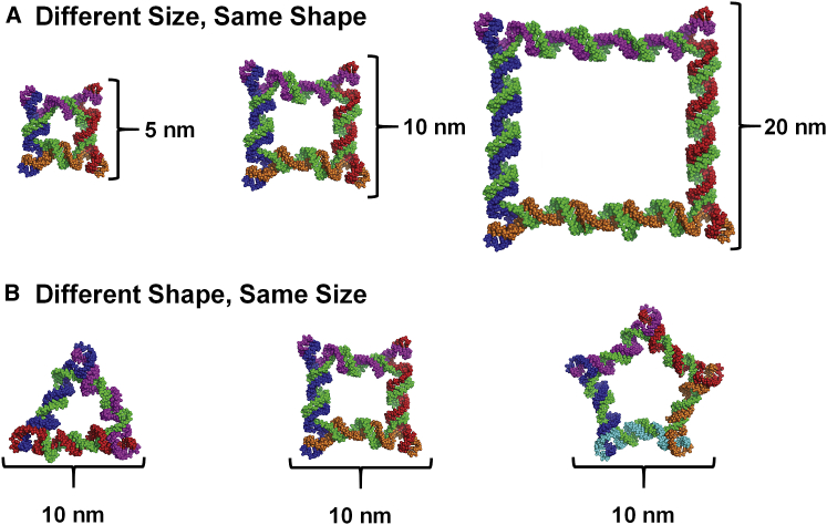
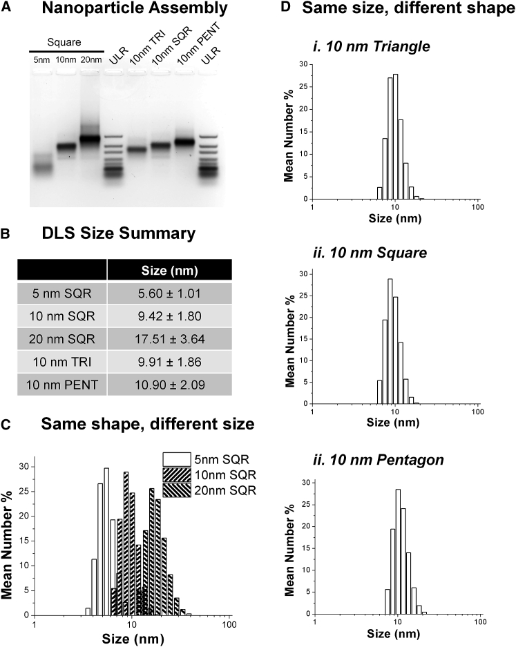
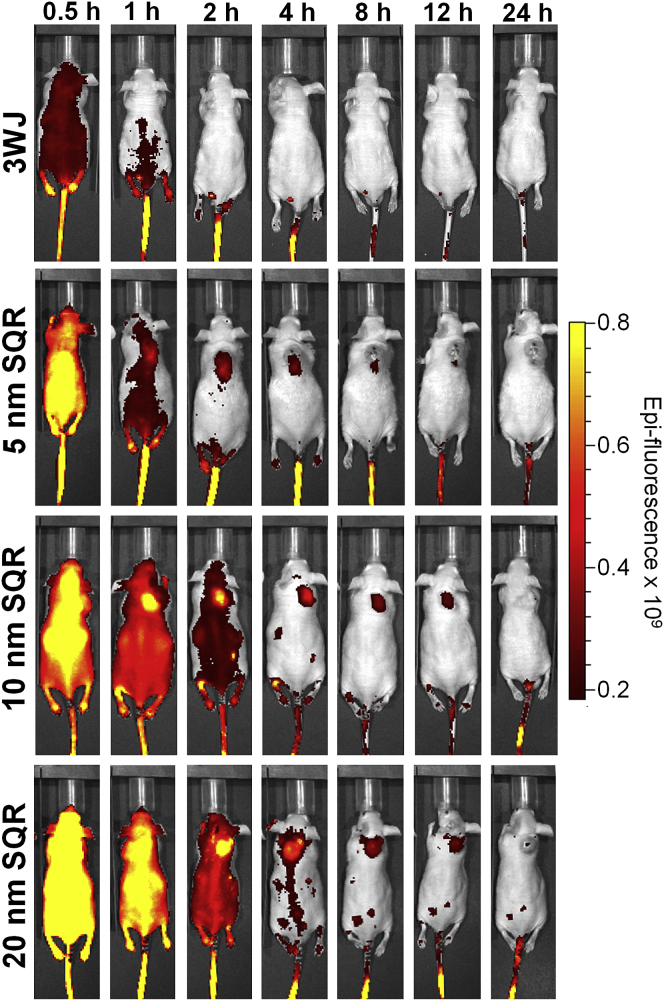
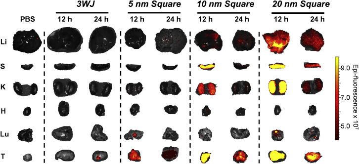
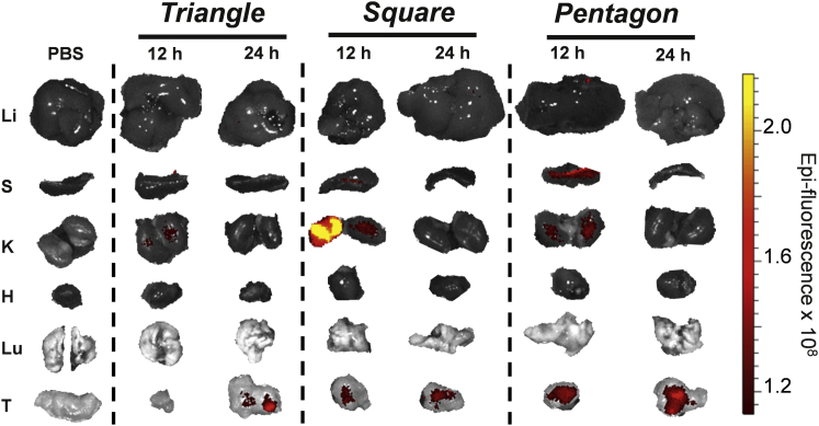
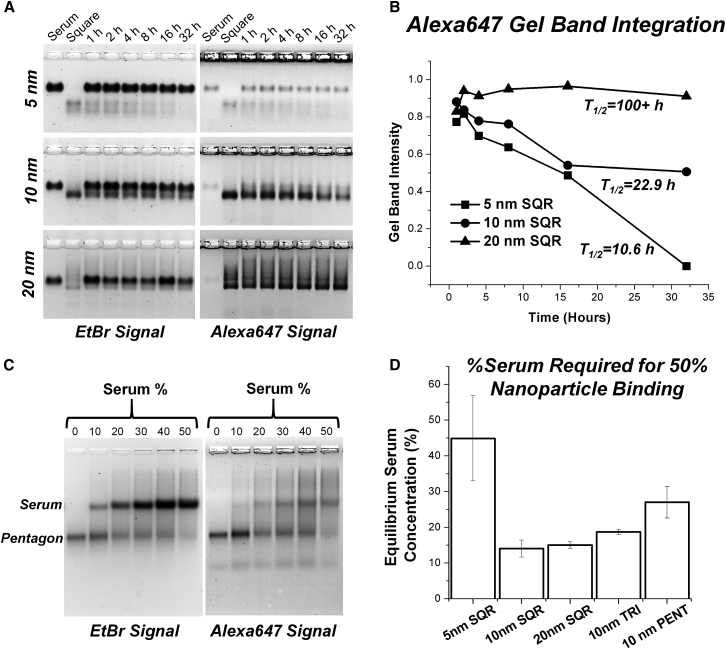
Drugs with ideal pharmacokinetic profile require long half-life but little organ accumulation. Generally, PK and organ accumulation are contradictory factors: smaller size leads to faster excretion and shorter half-lives and thus a lower tendency to reach targets; larger size leads to longer circulation but stronger organ accumulation that leads to toxicity. Organ accumulation has been reported to be size dependent due in large part to engulfing by macrophages. However, publications on the size effect are inconsistent because of complication by the effect of shape that varies from nanoparticle to nanoparticle. Unique to RNA nanotechnology, size could be tuned without a change in shape, resulting in a true size comparison. Here we investigated size effects using RNA squares of identical shape but varying size and shape effects using RNA triangles, squares, and pentagons of identical size but varying shape. We found that circulation time increased with increasing RNA nanoparticle size from 5-25 nm, which is the common size range of therapeutic RNA nanoparticles. Most particles were cleared from the body within 2 hr after systemic injection. Undetectable organ accumulation was found at any time for 5 nm particles. For 20 nm particles, weak signal was found after 24 hr, while accumulation in tumor was strongest during the entire study.
Keywords: RNA Nanotechnology; RNA nanoparticles; RNA nanostructure; bacteriophage phi29; motor pRNA; nanobiotechnology; pRNA 3WJ motif; phi29 DNA packaging motor; phi29 pRNA; viral DNA packaging.
Copyright © 2017 The American Society of Gene and Cell Therapy. Published by Elsevier Inc. All rights reserved.
Figures
Similar articles
-
Using Planar Phi29 pRNA Three-Way Junction to Control Size and Shape of RNA Nanoparticles for Biodistribution Profiling in Mice.Methods Mol Biol. 2017;1632:359-380. doi: 10.1007/978-1-4939-7138-1_23. Methods Mol Biol. 2017. PMID: 28730451
-
Favorable biodistribution, specific targeting and conditional endosomal escape of RNA nanoparticles in cancer therapy.Cancer Lett. 2018 Feb 1;414:57-70. doi: 10.1016/j.canlet.2017.09.043. Epub 2017 Oct 5. Cancer Lett. 2018. PMID: 28987384 Free PMC article. Review.
-
RNA nanoparticle distribution and clearance in the eye after subconjunctival injection with and without thermosensitive hydrogels.J Control Release. 2018 Jan 28;270:14-22. doi: 10.1016/j.jconrel.2017.11.028. Epub 2017 Nov 21. J Control Release. 2018. PMID: 29170141 Free PMC article.
-
Hydrophobic Effect from Conjugated Chemicals or Drugs on In Vivo Biodistribution of RNA Nanoparticles.Hum Gene Ther. 2018 Jan;29(1):77-86. doi: 10.1089/hum.2017.054. Epub 2017 Oct 12. Hum Gene Ther. 2018. PMID: 28557574 Free PMC article.
-
Methods for construction and characterization of simple or special multifunctional RNA nanoparticles based on the 3WJ of phi29 DNA packaging motor.Methods. 2018 Jul 1;143:121-133. doi: 10.1016/j.ymeth.2018.02.025. Epub 2018 Mar 9. Methods. 2018. PMID: 29530505 Free PMC article. Review.
Cited by
-
Recent strategies for enhanced delivery of mRNA to the lungs.Nanomedicine (Lond). 2025 May;20(9):1043-1069. doi: 10.1080/17435889.2025.2485669. Epub 2025 Apr 7. Nanomedicine (Lond). 2025. PMID: 40190037 Review.
-
Nucleic acid nanostructures for in vivo applications: The influence of morphology on biological fate.Appl Phys Rev. 2023 Mar;10(1):011304. doi: 10.1063/5.0121820. Appl Phys Rev. 2023. PMID: 36874908 Free PMC article. Review.
-
Nanodelivery of nucleic acids.Nat Rev Methods Primers. 2022;2:24. doi: 10.1038/s43586-022-00104-y. Epub 2022 Apr 14. Nat Rev Methods Primers. 2022. PMID: 35480987 Free PMC article.
-
RNA Nanoparticles as Rubber for Compelling Vessel Extravasation to Enhance Tumor Targeting and for Fast Renal Excretion to Reduce Toxicity.ACS Nano. 2020 Oct 27;14(10):13180-13191. doi: 10.1021/acsnano.0c04863. Epub 2020 Sep 16. ACS Nano. 2020. PMID: 32902260 Free PMC article.
-
The dynamic, motile and deformative properties of RNA nanoparticles facilitate the third milestone of drug development.Adv Drug Deliv Rev. 2022 Jul;186:114316. doi: 10.1016/j.addr.2022.114316. Epub 2022 May 5. Adv Drug Deliv Rev. 2022. PMID: 35526663 Free PMC article. Review.
References
-
- Duncan R. The dawning era of polymer therapeutics. Nat. Rev. Drug Discov. 2003;2:347–360. - PubMed
-
- Ferrari M. Cancer nanotechnology: opportunities and challenges. Nat. Rev. Cancer. 2005;5:161–171. - PubMed
-
- Quintanar-Guerrero D., Allémann E., Fessi H., Doelker E. Preparation techniques and mechanisms of formation of biodegradable nanoparticles from preformed polymers. Drug Dev. Ind. Pharm. 1998;24:1113–1128. - PubMed
-
- Banerjee R., Parida S., Maiti C., Mandal M., Dhara D. pH-degradable and thermoresponsive water-soluble core cross-linked polymeric nanoparticles as potential drug delivery vehicle for doxorubicin. RSC Advances. 2015;5:83565–83575.
-
- Lv S., Tang Z., Zhang D., Song W., Li M., Lin J., Liu H., Chen X. Well-defined polymer-drug conjugate engineered with redox and pH-sensitive release mechanism for efficient delivery of paclitaxel. J. Control. Release. 2014;194:220–227. - PubMed
Publication types
MeSH terms
Substances
Grants and funding
LinkOut - more resources
Full Text Sources
Other Literature Sources
