. 2018 Feb 5;8(1):2442.
doi: 10.1038/s41598-018-20866-4.

Selective Inhibition of Succinate Dehydrogenase in Reperfused Myocardium with Intracoronary Malonate Reduces Infarct Size

Affiliations

Selective Inhibition of Succinate Dehydrogenase in Reperfused Myocardium with Intracoronary Malonate Reduces Infarct Size

Laura Valls-Lacalle et al. Sci Rep. .

Erratum in

Abstract

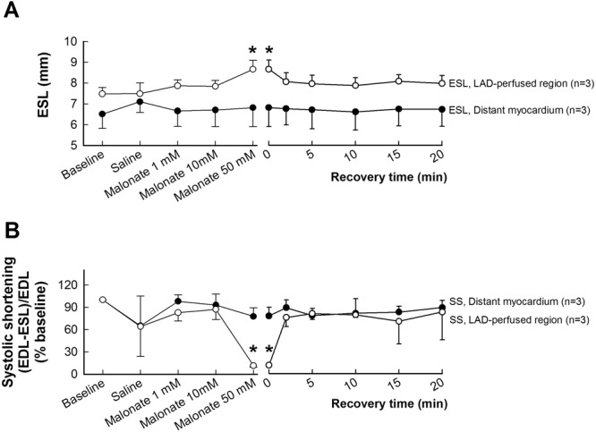
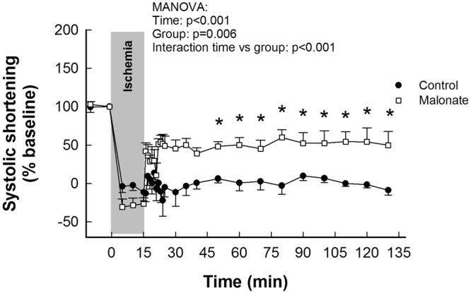
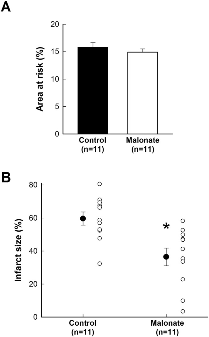
Inhibition of succinate dehydrogenase (SDH) with malonate during reperfusion reduces infarct size in isolated mice hearts submitted to global ischemia. However, malonate has toxic effects that preclude its systemic administration in animals. Here we investigated the effect of intracoronary malonate on infarct size in pigs submitted to transient coronary occlusion. Under baseline conditions, 50 mmol/L of intracoronary disodium malonate, but not lower concentrations, transiently reduced systolic segment shortening in the region perfused by the left anterior descending coronary artery (LAD) in open-chest pigs. To assess the effects of SDH inhibition on reperfusion injury, saline or malonate 10 mmol/L were selectively infused into the area at risk in 38 animals submitted to ischemia-reperfusion. Malonate improved systolic shortening in the area at risk two hours after 15 min of ischemia (0.18 ± 0.07 vs 0.00 ± 0.01 a.u., p = 0.025, n = 3). In animals submitted to 40 min of ischemia, malonate reduced reactive oxygen species production (MitoSOX staining) during initial reperfusion and limited infarct size (36.46 ± 5.35 vs 59.62 ± 4.00%, p = 0.002, n = 11), without modifying reperfusion arrhythmias. In conclusion, inhibition of SDH with intracoronary malonate during early reperfusion limits reperfusion injury and infarct size in pigs submitted to transient coronary occlusion without modifying reperfusion arrhythmias or contractile function in distant myocardium.


Conflict of interest statement

The authors declare that they have no competing interests.

Figures

Figure 1
Figure 1
Effects of succinate dehydrogenase inhibition with malonate in myocardial function in the LAD-perfused region in pigs under baseline conditions. Changes in end-systolic length (ESL) (A) and systolic segment shortening (SS, expressed as percentage of baseline value) (B), measured by ultrasonic crystals placed in the distant myocardium and in the LAD-perfused area, during intracoronary infusion of saline or increasing concentrations of disodium malonate in pigs. Baseline SS values were 0.21 ± 0.04 in distant myocardium and 0.13 ± 0.03 in the LAD-perfused region. *(p < 0.05) indicates significant differences vs. the corresponding baseline value.
Figure 2
Figure 2
Nuclear magnetic resonance spectroscopy analysis of porcine plasma samples obtained from pigs treated with malonate under baseline conditions. (A) Representative 1H-NMR spectra from porcine plasma samples obtained after intracoronary saline infusion or after treatment with malonate 1, 10 or 50 mmol/L. (B) Plasma concentrations of malonate under each condition. *(p < 0.05) indicates significant differences vs. values obtained after saline infusion.
Figure 3
Figure 3
Myocardial stunning in the area at risk in pigs submitted to transient coronary occlusion and treated with intracoronary malonate during initial reperfusion. Changes in systolic segment shortening (SS, expressed as percentage of baseline value) in the area at risk in pigs submitted to 15 min LAD coronary artery occlusion followed by reperfusion, and treated with intracoronary saline or 10 mmol/L of disodium malonate. Baseline SS values were 0.29 ± 0.03 for control animals and 0.31 ± 0.06 for malonate-treated pigs. *(p < 0.05) indicates significant differences vs. the corresponding value in the control group.
Figure 4
Figure 4
Effects of succinate dehydrogenase inhibition with intracoronary malonate during reperfusion on infarct size in pigs submitted to transient coronary occlusion. Size of the area at risk, expressed as percentage of the ventricular weight (A), in pigs submitted to 40 min of LAD coronary occlusion followed by 120 min of reperfusion, treated during initial reperfusion with an intracoronary infusion of saline (control) or disodium malonate 10 mmol/L. (B) Infarct size, expressed as percentage of the area at risk, in the same groups of animals. *(p < 0.05) indicates significant differences vs. control hearts (n = 11/group).
Figure 5
Figure 5
Incidence of reperfusion arrhythmias in pigs submitted to transient coronary occlusion and treated with intracoronary malonate during initial reperfusion. Total number of reperfusion ventricular tachycardias (VT) (A) and VT duration (B) in pigs submitted to 40 min of LAD coronary occlusion followed by 120 min of reperfusion, treated during initial reperfusion with an intracoronary infusion of saline (control) or disodium malonate 10 mmol/L. (C) Incidence of sustained ventricular tachycardia (VT, closed cercles) and ventricular fibrillation (VF, red squares) in the same group of animals. Blue square represents the duration of infusion.
Figure 6
Figure 6
Malonate, given during reperfusion, reduces myocardial ROS production in porcine tissue samples. Figures show representative confocal images depicting MitoSOX staining (in red) (excitation wavelength 488 nm, emission wavelength 520 nm) in myocardial biopsies obtained 5 min after flow restoration, from both the area at risk and control region, in pigs treated with intracoronary infusion of saline (controls) or malonate 10 mmol/L. Nuclei are stained in blue.

References

    1. Granger DN, Kvietys PR. Reperfusion injury and reactive oxygen species: The evolution of a concept. Redox. Biol. 2015;6:524–551. - PMC - PubMed
    1. Halestrap AP, Richardson AP. The mitochondrial permeability transition: a current perspective on its identity and role in ischaemia/reperfusion injury. J Mol Cell Cardiol. 2015;78:129–141. - PubMed
    1. Garcia-Dorado D, Ruiz-Meana M, Inserte J, Rodriguez-Sinovas A, Piper HM. Calcium-mediated cell death during myocardial reperfusion. Cardiovasc Res. 2012;94:168–180. - PubMed
    1. Jasova, M., Kancirova, I., Waczulikova, I. & Ferko, M. Mitochondria as a target of cardioprotection in models of preconditioning. J. Bioenerg. Biomembr. 10.1007/s10863-017-9720-1 (2017). - PubMed
    1. Liochev SI. Reactive oxygen species and the free radical theory of aging. Free Radic. Biol. Med. 2013;60:1–4. - PubMed

Publication types

MeSH terms