Review
. 2018 Apr;21(4):463-473.
doi: 10.1038/s41593-018-0080-x. Epub 2018 Feb 5.

Imbalance between firing homeostasis and synaptic plasticity drives early-phase Alzheimer's disease

Affiliations
Review

Imbalance between firing homeostasis and synaptic plasticity drives early-phase Alzheimer's disease

Boaz Styr et al. Nat Neurosci. 2018 Apr.

Abstract

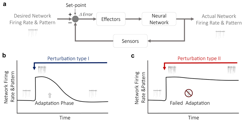
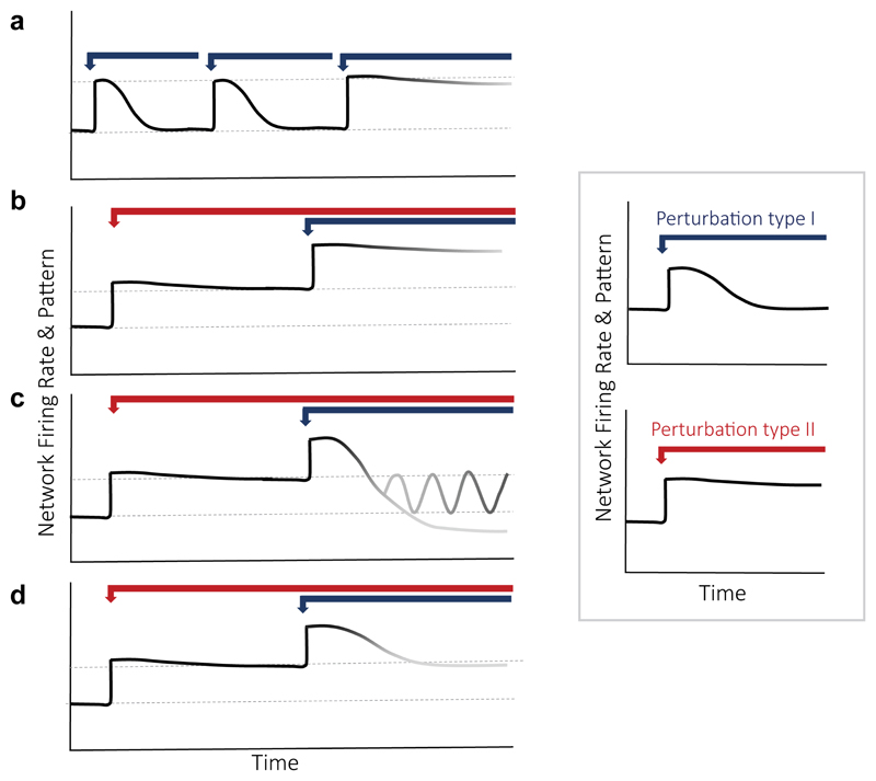
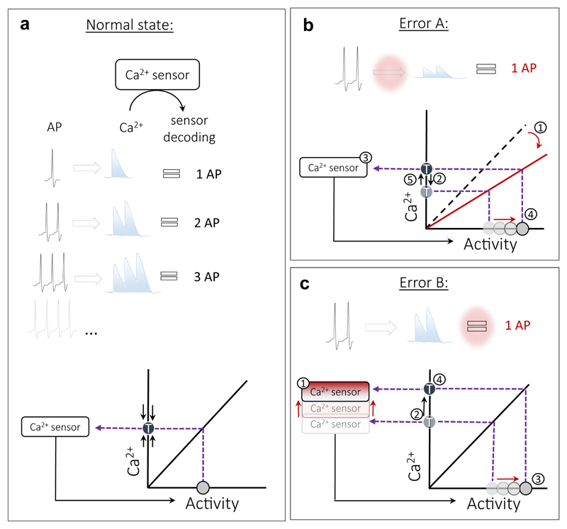
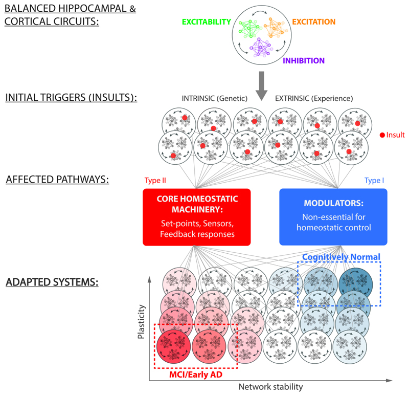
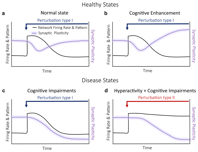
During recent years, the preclinical stage of Alzheimer's disease (AD) has become a major focus of research. Continued failures in clinical trials and the realization that early intervention may offer better therapeutic outcome triggered a conceptual shift from late-stage AD pathology to early-stage pathophysiology. While much effort has been directed at understanding the factors initiating AD, little is known about the principle basis underlying the disease progression at its early stages. In this Perspective, we suggest a hypothesis to explain the transition from 'silent' signatures of aberrant neural circuit activity to clinically evident memory impairments. Namely, we propose that failures in firing homeostasis and imbalance between firing stability and synaptic plasticity in cortico-hippocampal circuits represent the driving force of early disease progression. We analyze the main types of possible homeostatic failures and provide the essential conceptual framework for examining the causal link between dysregulation of firing homeostasis, aberrant neural circuit activity and memory-related plasticity impairments associated with early AD.


Figures

Figure 1
Figure 1. Firing homeostasis and its failure.
(a) A classic scheme of a homeostatic controller. In this case, the output of the network is the mean firing rate that is monitored by sensors and maintained at a set-point value by negative feedback mechanisms mediated via effectors. Any deviation from the desired firing rate is sensed as the difference between the desired output (the set-point) and the actual output. The error signal is then corrected via the activity of effectors. (b) Monitoring the activity of the same neurons for a long time enables to test if the mean firing rate in the network is stable. When a constant perturbation is introduced to elevate firing rates (blue arrow), homeostatic mechanisms are activated to adapt the system to the perturbation (adaptation phase). This type I perturbation relates to changes in non-essential, regulatory homeostatic components. It induces compensatory mechanisms that re-normalize firing rates, despite of the continued interference. (c) Under pathological conditions (perturbation type II, red arrow), homeostatic mechanisms fail to re-normalize firing rates, leaving the network in a hyperactive state due maladaptive responses. Type II perturbation relates to impairments of the core homeostatic machinery.
Figure 2
Figure 2. Experimental framework to investigate firing homeostasis failures.
Here we aim to investigate the effect of impairing core homeostatic machinery (perturbation type II) on firing stabilization following hyperactivity. (a) Accumulation of insults: A system that suffers multiple type 1 insults may initially be able to compensate, while it may eventually fail due to a restriction of the solution space following new insults. (b) Regulation is abolished: In this case, when type I is introduced in the presence of type II perturbation, the network does not compensate for the change in firing. This indicates type II restricts type I-induced homeostatic mechanisms and abolishes regulation of firing rates. (c) Regulation fails to reach the set-point: In the more complicated scenario, the network may overshoot, for example under malfunctioning of error signal estimations. The network may also enter an oscillation state if the kinetics of compensatory mechanisms is altered by type II perturbation. (d) Set-point is changed: In this example, when type I perturbation is introduced, homeostatic compensation mechanisms are still active, yet they trigger a compensation to the new steady-state level that type II perturbation imposed, indicating that type II affects firing set-point establishment. Inset: Perturbation type I (blue arrow), acutely augmenting spiking activity without impairing the essential elements of homeostatic system, induces homeostatic compensatory mechanisms that re-normalize firing rates to a set-point level (top panel). Perturbation type II (red arrow) affects mean firing rates without inducing a compensatory homeostatic response (bottom panel), indicating that type II is involved in regulation of firing rate stability.
Figure 3
Figure 3. Decoupling of Ca2+ sensors from spiking activity / stability.
(a) Coupling of spiking activity to Ca2+ sensor under physiological conditions. Top: Spiking activity produces changes in a sensed factor (Ca+2 for example). A sensor detects changes in Ca+2 and ‘translates’ spike-evoked Ca+2 transients to downstream effectors that then regulate spiking activity according to this information. Bottom: An example of a linear transfer function linking spiking activity to Ca+2 levels. The sensor corrects any deviation from the target Ca+2 level (T) by adjusting spiking activity. (b) In a pathological setting, the same spiking activity may produce less Ca2+, changing the slope of a transfer function. Ca2+ levels drop (1) even though spiking levels remain the same (2). The activated sensor (3) elevates spiking activity (4) in order to maintain the target Ca2+ levels (5), leading to a new hyperactive steady-state. (c) In another pathological setting, the sensitivity of the Ca+2 sensor to Ca2+ is reduced (1), shifting the target Ca2+ levels upwards (2). Excessive spiking activity is then produced (3) to maintain the new higher target Ca2+ levels (4).
Figure 4
Figure 4. FHP hypothesis: possible transitions from normal to early AD states.
A fully functional homeostatic controller enables a balance between excitatory synaptic drive (excitation), inhibitory synaptic drive (inhibition) and intrinsic excitability. Genetic, pharmacological and experience-dependent life events can trigger malfunction at a particular node (red dot) in the network, affecting firing stability. Depending on the initial state of the regulatory system and the type of insult inflicted, a subset of solutions become maladaptive, resulting in cognitive impairments at the early AD stages, while the majority retain normal cognitive function. According to the FHP hypothesis, the insults that impair the core homeostatic machinery reduce the homeostatic capacity of the network and lead to a spectrum of maladaptive responses, resulting in early AD. Within the AD subset of solutions, not all have the same functional features. Some might manifest it hyperactivity, while others might lead to impaired plasticity, and these dysfunctions may extensively overlap. On the other side of the spectrum are insults affecting mechanisms that are non-essential for homeostatic response. These lead to a spectrum of adaptive solutions that enable functional re-normalization and preserve cognitive function.
Figure 5
Figure 5. Balance of firing stability – synaptic plasticity and its disruption in early AD stages.
(a) Normal healthy state: A perturbation type I results in a transient increase in firing rates (black line) with concomitant reduction in synaptic plasticity (purple line). The adaptive mechanisms induced by a perturbation result in re-normalization of both, firing rates and synaptic plasticity. (b) Cognitive enhancement: Adaptive mechanisms induced by a perturbation type I to re-normalize firing rates, increase some types of synaptic plasticity. An example: a decrease in release probability in response to hyperactivity, resulting in increase of synaptic facilitation. (c) Cognitive impairments: Adaptive mechanisms to perturbation type I cause reduction in synaptic plasticity as the price for firing stability. (d) Cognitive impairments: Adaptive mechanisms to perturbation type II cause hyperactivity with subsequent reduction in synaptic plasticity.

References

    1. Hardy J, Selkoe DJ. The amyloid hypothesis of Alzheimer's disease: progress and problems on the road to therapeutics. Science. 2002;297:353–356. - PubMed
    1. Selkoe DJ, Hardy J. The amyloid hypothesis of Alzheimer's disease at 25 years. EMBO Molecular Medicine. 2016;8:595–608. - PMC - PubMed
    1. Small SA, Duff K. Linking Aβ and Tau in Late-Onset Alzheimer's Disease: A Dual Pathway Hypothesis. Neuron. 2008;60:534–542. - PMC - PubMed
    1. Herrup K. The case for rejecting the amyloid cascade hypothesis. Nat Neurosci. 2015:794–799. - PubMed
    1. De Strooper B, Karran E. The Cellular Phase of Alzheimer’s Disease. Cell. 2016;164:603–615. - PubMed

Publication types