"Soft that molds the hard:" Geometric morphometry of lateral atlantoaxial joints focusing on the role of cartilage in changing the contour of bony articular surfaces
- PMID: 29403249
- PMCID: PMC5763594
- DOI: 10.4103/jcvjs.JCVJS_109_17
"Soft that molds the hard:" Geometric morphometry of lateral atlantoaxial joints focusing on the role of cartilage in changing the contour of bony articular surfaces
Abstract
Purpose: The existing literature on lateral atlantoaxial joints is predominantly on bony facets and is unable to explain various C1-2 motions observed. Geometric morphometry of facets would help us in understanding the role of cartilages in C1-2 biomechanics/kinematics.
Objective: Anthropometric measurements (bone and cartilage) of the atlantoaxial joint and to assess the role of cartilages in joint biomechanics.
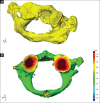
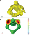
Materials and methods: The authors studied 10 cadaveric atlantoaxial lateral joints with the articular cartilage in situ and after removing it, using three-dimensional laser scanner. The data were compared using geometric morphometry with emphasis on surface contours of articulating surfaces.
Results: The bony inferior articular facet of atlas is concave in both sagittal and coronal plane. The bony superior articular facet of axis is convex in sagittal plane and is concave (laterally) and convex medially in the coronal plane. The bony articulating surfaces were nonconcordant. The articular cartilages of both C1 and C2 are biconvex in both planes and are thicker than the concavities of bony articulating surfaces.
Conclusion: The biconvex structure of cartilage converts the surface morphology of C1-C2 bony facets from concave on concavo-convex to convex on convex. This reduces the contact point making the six degrees of freedom of motion possible and also makes the joint gyroscopic.
Keywords: Articular surface; atlantoaxial joint; atlas; axis; cartilage; three-dimensional morphometry.
Conflict of interest statement
There are no conflicts of interest.
Figures


References
-
- Dickman CA, Theodore N, Crawford NR. Biomechanics of the craniovertebral junction. In: Bambakidis NC, Dickman CA, Spetzler RF, Sonntag VK, editors. Surgery of the Craniovertebral Junction. 2nd ed. New York, USA: Thieme; 2013. pp. 52–62.
-
- Goel A, Kulkarni AG, Sharma P. Reduction of fixed atlantoaxial dislocation in 24 cases: Technical note. J Neurosurg Spine. 2005;2:505–9. - PubMed
-
- Salunke P, Sahoo SK, Deepak AN, Ghuman MS, Khandelwal NK. Comprehensive drilling of the C1-2 facets to achieve direct posterior reduction in irreducible atlantoaxial dislocation. J Neurosurg Spine. 2015;23:294–302. - PubMed
-
- Salunke P, Sahoo S, Khandelwal NK, Ghuman MS. Technique for direct posterior reduction in irreducible atlantoaxial dislocation: Multi-planar realignment of C1-2. Clin Neurol Neurosurg. 2015;131:47–53. - PubMed
-
- Goel A. Treatment of basilar invagination by atlantoaxial joint distraction and direct lateral mass fixation. J Neurosurg Spine. 2004;1:281–6. - PubMed
LinkOut - more resources
Full Text Sources
Other Literature Sources
Miscellaneous

