. 2017 Sep 18:10:1179064417730559.
doi: 10.1177/1179064417730559. eCollection 2017.

Upregulated Heat Shock Proteins After Hyperthermic Chemotherapy Point to Induced Cell Survival Mechanisms in Affected Tumor Cells From Peritoneal Carcinomatosis

Affiliations

Upregulated Heat Shock Proteins After Hyperthermic Chemotherapy Point to Induced Cell Survival Mechanisms in Affected Tumor Cells From Peritoneal Carcinomatosis

Tanja Grimmig et al. Cancer Growth Metastasis. .

Abstract

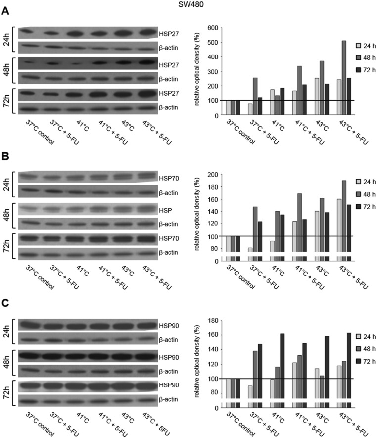
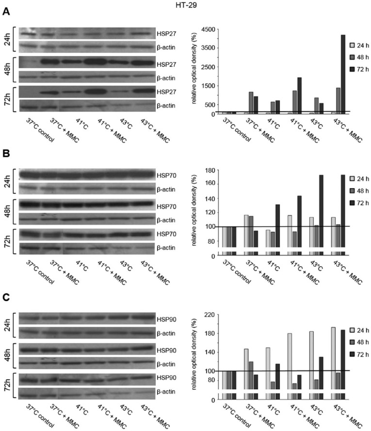
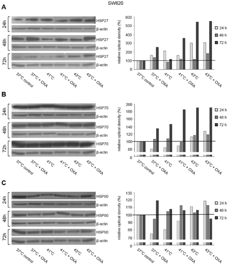
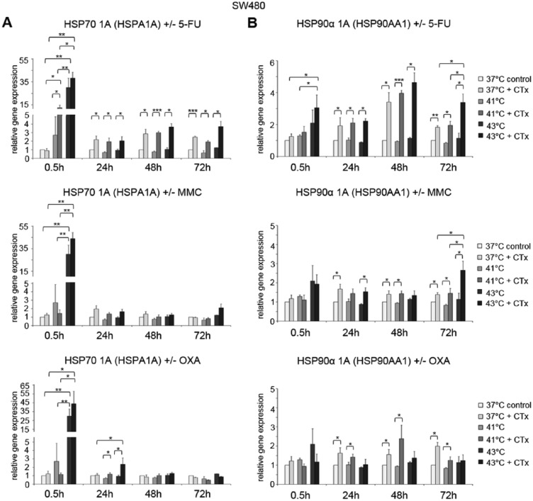
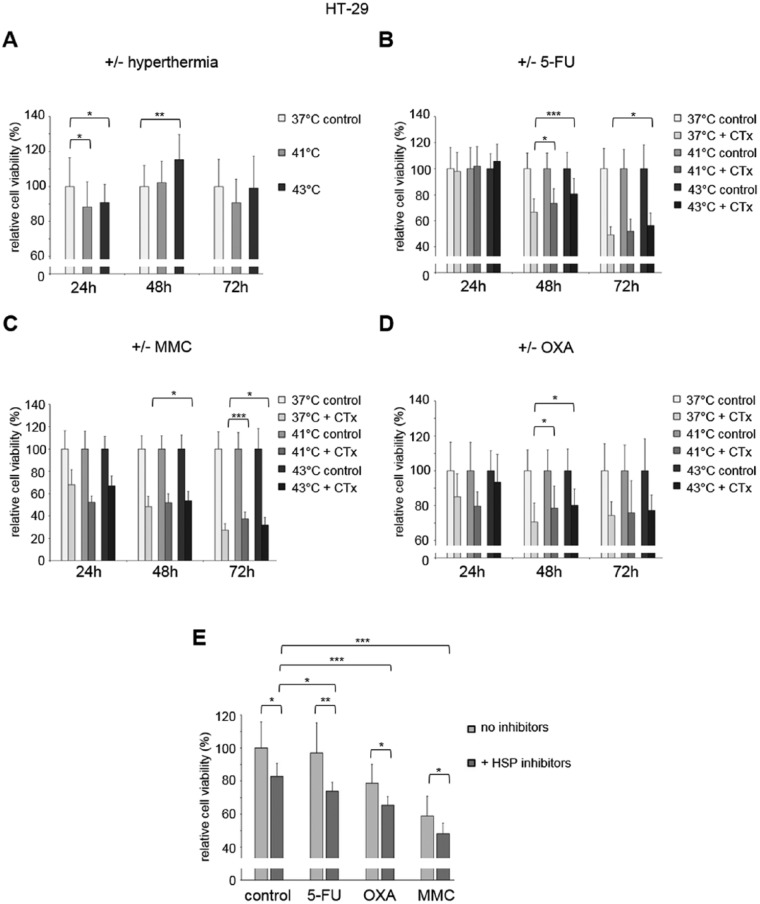
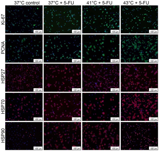
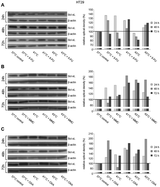
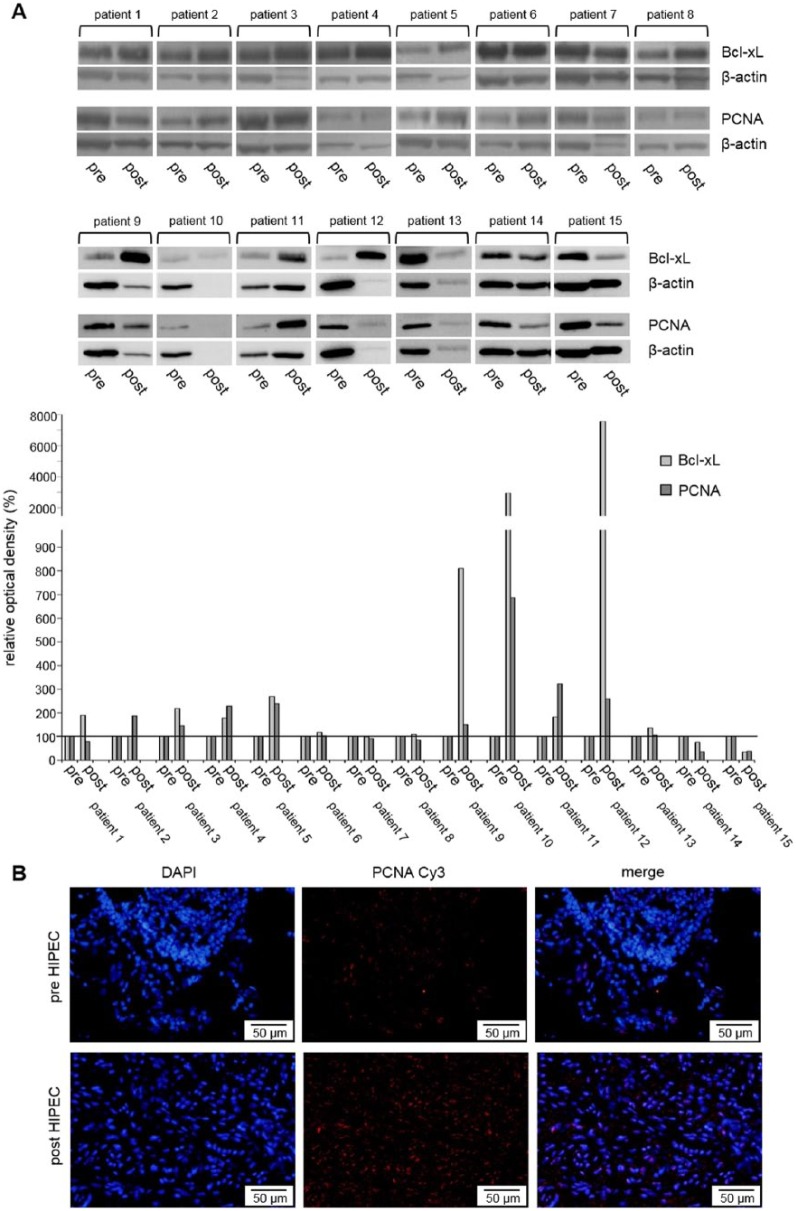
In patients with peritoneal carcinomatosis cytoreductive surgery combined with hyperthermic intraperitoneal chemotherapy (HIPEC) represents a promising treatment strategy. Here, we studied the role of hyperthermic chemotherapy on heat shock protein (HSP) expression and induction of tumor cell death and survival. HSP27, HSP70, and HSP90 combined with effects on tumor cell proliferation and chemosensitivity were analyzed in human colon cancer. Hyperthermic chemotherapy resulted in significant HSP27/HSP70 and HSP90 gene/protein overexpression in analyzed HT-29/SW480/SW620 colon cancer cells and peritoneal metastases from patients displaying amplified expression of proliferation markers, proliferating cell nuclear antigen and antiapoptotic protein Bcl-xL. Moreover, functionally increased chemoresistance against 5-fluorouracil/mitomycin C and oxaliplatin after hyperthermic chemotherapy points to induced survival mechanisms in cancer cells. In conclusion, the results indicate that intracellular HSP-associated antiapoptotic and proliferative effects after hyperthermic chemotherapy negatively influence beneficial effects of hyperthermic chemotherapy-induced cell death. Therefore, blocking HSPs could be a promising strategy to further improve the rate of tumor cell death and outcome of patients undergoing HIPEC therapy.

Keywords: Heat shock proteins; apoptosis; chemoresistance; hyperthermia; hyperthermic intraperitoneal chemotherapy; peritoneal carcinomatosis.


Conflict of interest statement

Declaration of conflicting interests:The author(s) declared no potential conflicts of interest with respect to the research, authorship, and/or publication of this article.

Figures

Figure 1.
Figure 1.
Western blot analysis of (A) HSP27, (B) HSP70, and (C) HSP90 in SW480 cells 24, 48, and 72 hours after treatment (60 minutes) with hyperthermia including or without additional chemotherapy using 5-fluorouracil (5-FU). β-actin probe was used as a control for protein loading. Relative optical density was determined using ImageJ software: values for proteins of interest were calculated in relation to values of loading controls. Normothermic cells (37°C control) were standardized to baseline.
Figure 2.
Figure 2.
Western blot analysis of (A) HSP27, (B) HSP70, and (C) HSP90 in HT-29 cells 24, 48, and 72 hours after treatment (60 minutes) with hyperthermia including or without additional chemotherapy using mitomycin C (MMC). β-actin probe was used as a control for protein loading. Relative optical density was determined using ImageJ software: values for proteins of interest were calculated in relation to values of loading controls. Normothermic cells (37°C control) were standardized to baseline.
Figure 3.
Figure 3.
Western blot analysis of (A) HSP27, (B) HSP70, and (C) HSP90 in SW620 cells 24, 48, and 72 hours after treatment (60 minutes) with hyperthermia including or without additional chemotherapy using oxaliplatin (OXA). β-actin probe was used as a control for protein loading. Relative optical density was determined using ImageJ software: values for proteins of interest were calculated in relation to values of loading controls. Normothermic cells (37°C control) were standardized to baseline.
Figure 4.
Figure 4.
(A) Gene expression analysis (real-time polymerase chain reaction) of HSPA1A (HSP70 1A) and (B) HSP90AA1 (HSP90α 1A), in SW480 cells 0.5, 24, 48, and 72 hours after treatment (60 minutes) with hyperthermia including or without additional chemotherapy (CTx) using 5-fluorouracil (5-FU), mitomycin C (MMC), or oxaliplatin (OXA). Normothermic cells without cytostatic treatment (37°C control) were standardized to baseline. The relative gene expression is expressed as 2−ΔΔCq (= fold difference, FD). Reproducibility was confirmed by 3 independent runs. *P < .05; **P < .0005; ***P < .0001 (Student t test).
Figure 5.
Figure 5.
Gene expression analysis (real-time polymerase chain reaction) of HSPB1 (HSP27 1) in HT-29 cells 0.5, 24, 48, and 72 hours after treatment (60 minutes) with hyperthermia including or without additional chemotherapy (CTx) using 5-fluorouracil (5-FU), mitomycin C (MMC), or oxaliplatin (OXA). Normothermic cells without cytostatic treatment (37°C control) were standardized to baseline. The relative gene expression is expressed as 2−ΔΔCq (= fold difference, FD). Reproducibility was confirmed by 3 independent runs. *P < .05; **P < .0005; ***P < .0001 (Student t test).
Figure 6.
Figure 6.
(A) MTS proliferation assay of HT-29 colon cancer cells 24, 48, and 72 hours after single hyperthermic treatment (60 minutes) or (B) additional chemotherapy (CTx) during hyperthermic intraperitoneal chemotherapy–like conditions using 5-fluorouracil (5-FU), (C) mitomycin C (MMC), or (D) oxaliplatin (OXA). (E) MTS proliferation assay of HT-29 colon cancer cells 24 hours after hyperthermic chemotherapy (43°C) including or without additional combined HSP70/90 inhibition using 17-AAG (HSP90) and VER155008 (HSP70). Colon cancer cells under normothermic condition (A, 37°C control), colon cancer cells without cytostatic treatment (B-D, control), and colon cancer cells without exposure to cytostatic agents and HSP inhibitors (E, control), respectively, were standardized to baseline. *P < .05; **P < .0005; ***P < .0001 (Student t test).
Figure 7.
Figure 7.
Gene expression analysis (real-time polymerase chain reaction) of proliferation markers (A) PCNA (proliferating cell nuclear antigen and (B) Ki-67 in HT-29 cells 0.5, 24, 48, and 72 hours after treatment (60 minutes) with hyperthermia including or without additional chemotherapy (CTx) using 5-fluorouracil (5-FU), mitomycin C (MMC), or oxaliplatin (OXA). Colon cancer cells under normothermic condition without cytostatic treatment (37°C control) were standardized to baseline. The relative gene expression is expressed as 2−ΔΔCq (= fold difference, FD). Reproducibility was confirmed by 3 independent runs. *P < .05; **P < .0005; ***P < .0001 (Student t test).
Figure 8.
Figure 8.
Immunofluorescent stainings of PCNA (green), Ki-67 (green), HSP27 (red), HSP70 (red), and HSP90 (red). FITC (fluorescein isothiocyanate), green; Cy3 (indocarbocyanine), red; DAPI (4′,6-diamidino-2-phenylindole dihydrochloride); blue—nuclear counterstaining.
Figure 9.
Figure 9.
Western blot analysis of PCNA (proliferating cell nuclear antigen) in HT-29 cells 24, 48, and 72 hours after treatment (60 minutes) with (A) hyperthermia including or without additional chemotherapy using (B) 5-fluorouracil (5-FU), mitomycin C (MMC), or (C) oxaliplatin (OXA). β-actin probe was used as a control for protein loading. Relative optical density was determined using ImageJ software: values for proteins of interest were calculated in relation to values of loading controls. Normothermic cells without cytostatic treatment (37°C control) were standardized to baseline.
Figure 10.
Figure 10.
Western blot analysis of antiapoptotic Bcl-xL in HT-29 cells 24, 48, and 72 hours after treatment (60 minutes) with hyperthermia including or without additional chemotherapy using (A) 5-fluorouracil (5-FU), (B) mitomycin C (MMC), or (C) oxaliplatin (OXA). β-actin probe was used as a control for protein loading. Relative optical density was determined using ImageJ software: values for proteins of interest were calculated in relation to values of loading controls. Normothermic cells without cytostatic treatment (37°C control) were standardized to baseline.
Figure 11.
Figure 11.
(A) Western blot analysis of antiapoptotic Bcl-xL and proliferation marker PCNA (proliferating cell nuclear antigen) in tumor tissue samples from peritoneal carcinomatosis of patients with colorectal cancer before (pre) and after (post) HIPEC. (B) Immunofluorescent staining of PCNA in peritoneal tumor tissue samples before (pre) and after (post) HIPEC. Cy3 (indocarbocyanine), red; DAPI (4′,6-diamidino-2-phenylindole dihydrochloride), blue—nuclear counterstaining. HIPEC indicates hyperthermic intraperitoneal chemotherapy. β-actin probe was used as a control for protein loading. Relative optical density was determined using ImageJ software: values for proteins of interest were calculated in relation to values of loading controls. Pre-HIPEC samples were standardized to baseline.

Similar articles

Cited by

References

    1. Elias D, Lefevre JH, Chevalier J, et al. Complete cytoreductive surgery plus intraperitoneal chemohyperthermia with oxaliplatin for peritoneal carcinomatosis of colorectal origin. J Clin Oncol. 2009;27:681–685. - PubMed
    1. Sugarbaker PH, Yonemura Y. Clinical pathway for the management of resectable gastric cancer with peritoneal seeding: best palliation with a ray of hope for cure. Oncology. 2000;58:96–107. - PubMed
    1. Franko J, Shi Q, Goldman CD, et al. Treatment of colorectal peritoneal carcinomatosis with systemic chemotherapy: a pooled analysis of north central cancer treatment group phase III trials N9741 and N9841. J Clin Oncol. 2012;30:263–267. - PMC - PubMed
    1. Spiliotis JD. Peritoneal carcinomatosis cytoreductive surgery and HIPEC: a ray of hope for cure. Hepatogastroenterology. 2010;57:1173–1177. - PubMed
    1. Haslinger M, Francescutti V, Attwood K, et al. A contemporary analysis of morbidity and outcomes in cytoreduction/hyperthermic intraperitoneal chemoperfusion. Cancer Med. 2013;2:334–342. - PMC - PubMed

LinkOut - more resources