

New Progress on the Role of Glia in Iron Metabolism and Iron-Induced Degeneration of Dopamine Neurons in Parkinson's Disease
- PMID: 29403352
- PMCID: PMC5780449
- DOI: 10.3389/fnmol.2017.00455
New Progress on the Role of Glia in Iron Metabolism and Iron-Induced Degeneration of Dopamine Neurons in Parkinson's Disease
Abstract
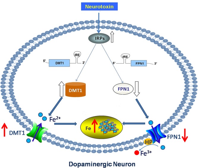
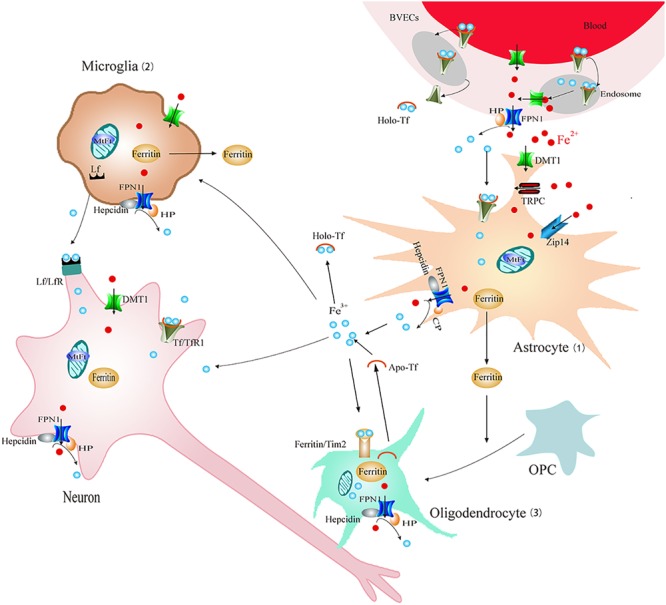
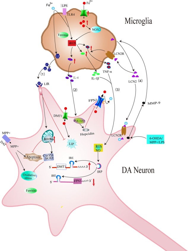
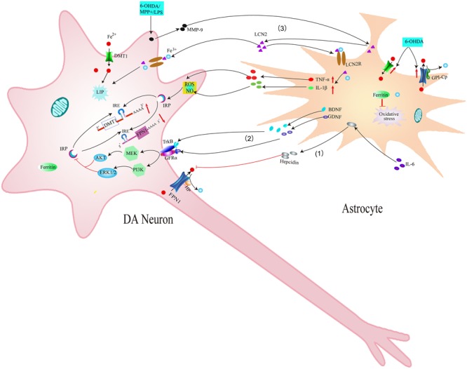
It is now increasingly appreciated that glial cells play a critical role in the regulation of iron homeostasis. Impairment of these properties might lead to dysfunction of iron metabolism and neurodegeneration of neurons. We have previously shown that dysfunction of glia could cause iron deposit and enhance iron-induced degeneration of dopamine (DA) neurons in Parkinson's disease (PD). There also has been a substantial growth of knowledge regarding the iron metabolism of glia and their effects on iron accumulation and degeneration of DA neurons in PD in recent years. Here, we attempt to describe the role of iron metabolism of glia and the effect of glia on iron accumulation and degeneration of DA neurons in the substantia nigra of PD. This could provide evidence to reveal the mechanisms underlying nigral iron accumulation of DA neurons in PD and provide the basis for discovering new potential therapeutic targets for PD.
Keywords: Parkinson’s disease; dopamine neurons; glia; iron; iron transporters.
Figures
Similar articles
-
[Glial cells are involved in iron accumulation and degeneration of dopamine neurons in Parkinson's disease].Sheng Li Xue Bao. 2016 Aug 25;68(4):455-63. Sheng Li Xue Bao. 2016. PMID: 27546505 Review. Chinese.
-
Proteasome inhibition modeling nigral neuron degeneration in Parkinson's disease.J Neurochem. 2010 Oct;115(1):188-99. doi: 10.1111/j.1471-4159.2010.06914.x. Epub 2010 Aug 19. J Neurochem. 2010. PMID: 20649845
-
The Therapeutic Implications of Tea Polyphenols Against Dopamine (DA) Neuron Degeneration in Parkinson's Disease (PD).Cells. 2019 Aug 16;8(8):911. doi: 10.3390/cells8080911. Cells. 2019. PMID: 31426448 Free PMC article.
-
The Association of Iron and the Pathologies of Parkinson's Diseases in MPTP/MPP+-Induced Neuronal Degeneration in Non-human Primates and in Cell Culture.Front Aging Neurosci. 2019 Aug 30;11:215. doi: 10.3389/fnagi.2019.00215. eCollection 2019. Front Aging Neurosci. 2019. PMID: 31543809 Free PMC article.
-
Role of iron in UPS impairment model of Parkinson's disease.Parkinsonism Relat Disord. 2014 Jan;20 Suppl 1:S158-61. doi: 10.1016/S1353-8020(13)70038-5. Parkinsonism Relat Disord. 2014. PMID: 24262171 Review.
Cited by
-
New Insights into the Role of Ferritin in Iron Homeostasis and Neurodegenerative Diseases.Mol Neurobiol. 2021 Jun;58(6):2812-2823. doi: 10.1007/s12035-020-02277-7. Epub 2021 Jan 28. Mol Neurobiol. 2021. PMID: 33507490 Review.
-
Iron and manganese-related CNS toxicity: mechanisms, diagnosis and treatment.Expert Rev Neurother. 2019 Mar;19(3):243-260. doi: 10.1080/14737175.2019.1581608. Epub 2019 Feb 21. Expert Rev Neurother. 2019. PMID: 30759034 Free PMC article. Review.
-
Pathogenesis of α-Synuclein in Parkinson's Disease: From a Neuron-Glia Crosstalk Perspective.Int J Mol Sci. 2022 Nov 25;23(23):14753. doi: 10.3390/ijms232314753. Int J Mol Sci. 2022. PMID: 36499080 Free PMC article. Review.
-
Parkinson Disease Signaling Pathways, Molecular Mechanisms, and Potential Therapeutic Strategies: A Comprehensive Review.Int J Mol Sci. 2025 Jul 3;26(13):6416. doi: 10.3390/ijms26136416. Int J Mol Sci. 2025. PMID: 40650193 Free PMC article. Review.
-
Iron dyshomeostasis and time-course changes in iron-uptake systems and ferritin level in relation to pro-inflammatory microglia polarization in sepsis-induced encephalopathy.Front Physiol. 2022 Aug 12;13:953206. doi: 10.3389/fphys.2022.953206. eCollection 2022. Front Physiol. 2022. PMID: 36035473 Free PMC article.
References
Publication types
LinkOut - more resources
Full Text Sources
Other Literature Sources