. 2018 Jan 17:8:2705.
doi: 10.3389/fmicb.2017.02705. eCollection 2017.

Lactobacillus rhamnosus GG Affects Microbiota and Suppresses Autophagy in the Intestines of Pigs Challenged with Salmonella Infantis

Affiliations

Lactobacillus rhamnosus GG Affects Microbiota and Suppresses Autophagy in the Intestines of Pigs Challenged with Salmonella Infantis

Wei Zhang et al. Front Microbiol. .

Abstract

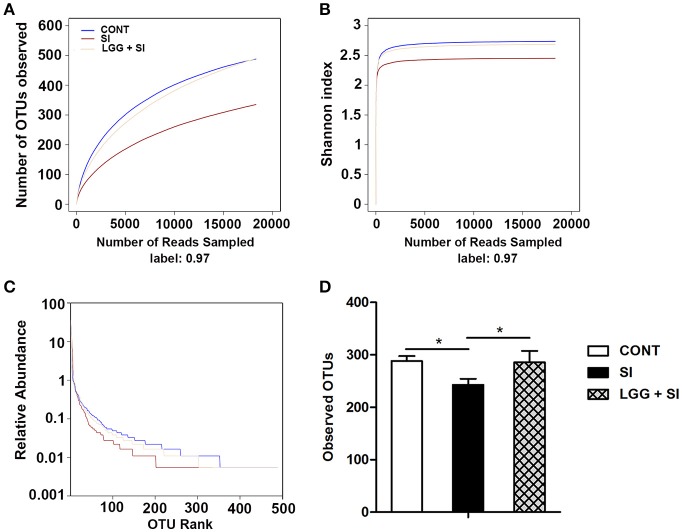
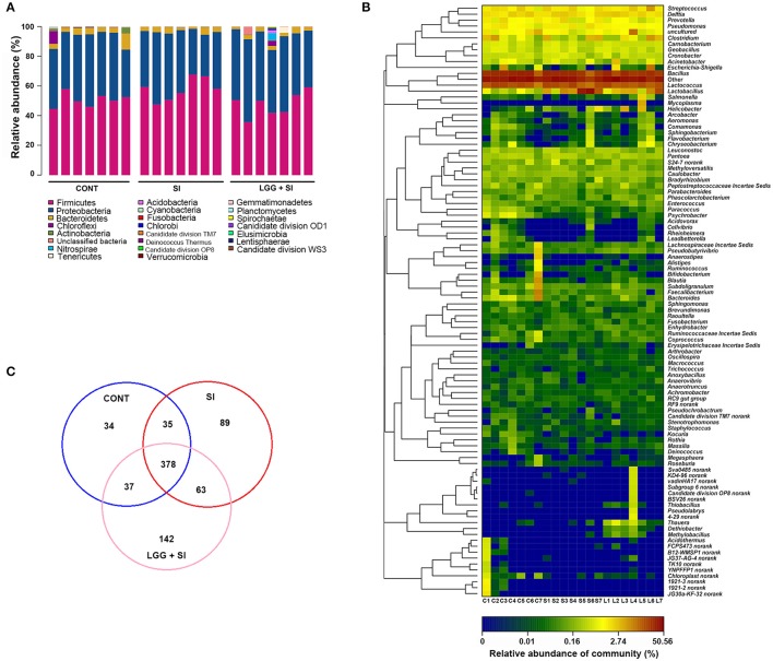
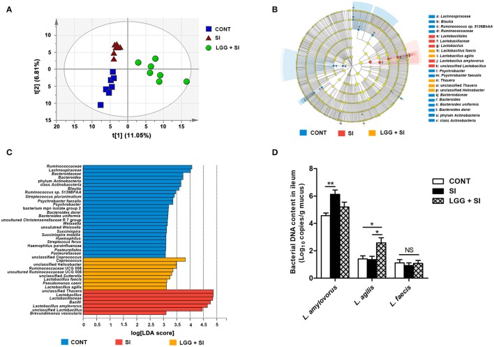
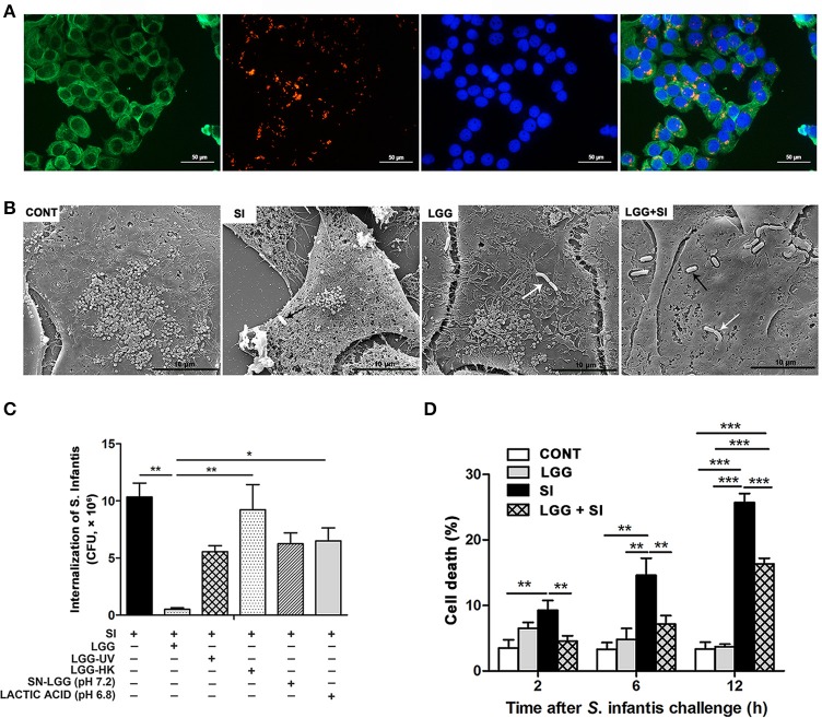
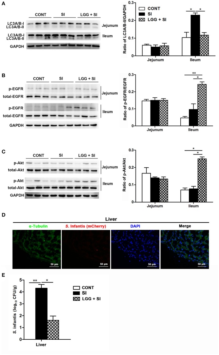
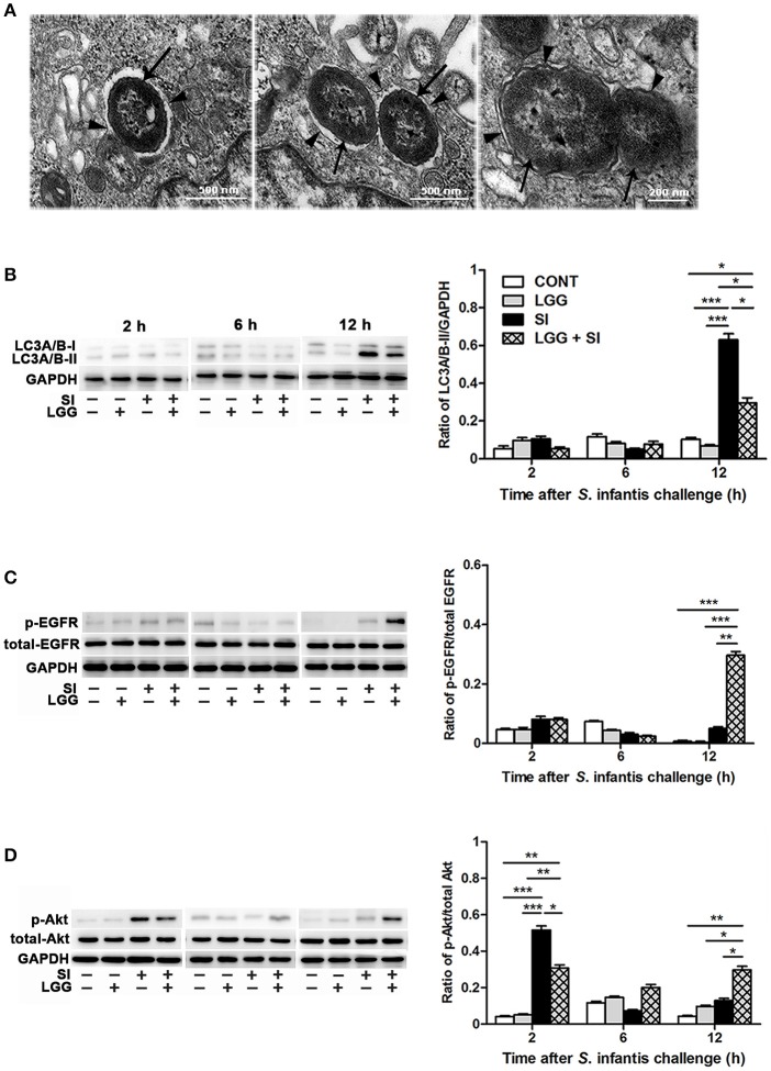
Salmonella enterica serovar Infantis (S. Infantis) is a common source of foodborne gastroenteritis worldwide. Here, Lactobacillus rhamnosus GG (LGG) was administrated to weaned piglets for 1 week before S. Infantis challenge. S. Infantis caused decreased ileal mucosal microbiota diversity, a dramatic Lactobacillus amylovorus bloom, and decreased abundance of Arsenicicoccus, Janibacter, Kocuria, Nocardioides, Devosia, Paracoccus, Psychrobacter, and Weissella. The beneficial effect of LGG correlated with the moderate expansion of L. amylovorus, L. agilis, and several members of the phyla Proteobacteria, Firmicutes, and Bacteroidetes. S. Infantis translocation to the liver was decreased in the LGG-pretreated piglets. An in vitro model of LGG and S. Infantis co-incubation (involving the porcine intestinal epithelial cell line IPEC-J2) was established, and nalidixic acid was used to kill the extracellular S. Infantis. LGG suppressed the initial S. Infantis invasion in the IPEC-J2 cells and deceased the rate of cell death. LGG inhibited S. Infantis-induced autophagy and promoted epidermal growth factor receptor (EGFR) and Akt phosphorylation in both the ileum and IPEC-J2 cells. Our findings suggest that LGG inhibited S. Infantis-induced autophagy by promoting EGFR-mediated activation of the negative mediator Akt, which, in turn, suppressed intestinal epithelial cell death and thus restricted systemic S. Infantis infection. LGG can restore the gut microbiota balance and preserve the autophagy-related intestinal epithelial barrier, thereby controlling infections.

Keywords: EGFR/Akt; Lactobacillus rhamnosus; Salmonella Infantis; autophagy; gut microbiota; pig.


Figures

Figure 1
Figure 1
Diversity analysis of ileal mucosal microbiota. The rarefaction abundance curves (A), Shannon diversity curves (B), and rank abundance curves (C) were used to estimate the ileal mucosal microbiota diversity at 97% similarity in pigs (n = 7 per group) that received oral sterile physiological saline (CONT), oral sterile physiological saline followed by S. Infantis (5 × 10¹⁰ CFU/ml, 10 ml) challenge (SI), or LGG (1 × 10⁹ CFU/ml, 10 ml/day) for 1 week followed by S. Infantis challenge (LGG+SI). (D) The difference in operational taxonomic units (OTUs) observed among the three groups was analyzed using one-way ANOVA (mean ± SEM, n = 7 per group). *P < 0.05.
Figure 2
Figure 2
Ileal mucosal microbiota profiles of S. Infantis-infected pigs pretreated with LGG. (A) Ileal mucosal microbiota profiles at the phylum level. The stacked bars show the combined relative abundance of phylum-level OTUs per pig. Colors are assigned for all phyla detected. (B) Heatmap showing the spatial distributions of genera, indicating the relative abundance of the top 100 genera per pig in the CONT (C), SI (S), and LGG+SI (L) groups. Genera are clustered to the left based on relative abundance. The relative abundance of each genus is indicated by a color gradient from blue (low abundance) to red (high abundance). The letters (C, S, or L) combined with single digits (1 to 7) represent the individual pigs in the corresponding groups. (C) Venn diagrams illustrating overlapping OTUs in the ileal mucosal microbiota of the three groups (n = 7 per group).
Figure 3
Figure 3
LGG pretreatment altered the ileal mucosal microbiota structure during S. Infantis challenge. (A) Two-dimensional partial least squares-discriminant analysis (PLS-DA) score plots based on the relative abundance of OTUs, showing the differences in the ileal mucosal microbiota in response to LGG pretreatment and S. Infantis challenge. LEfSe was performed to identify the most differentially abundant taxa in each of the three groups (n = 7 per group). (B) Taxonomic cladogram indicating the phylogenetic distribution of microbial lineages associated with the three groups (n = 7 per group). Lineages with an LDA value >3 are displayed. The diameter of each dot is proportional to its effect size. (C) Only taxa with an LDA value >3 are shown. (D) Quantitative PCR of bacterial 16S rRNA gene in ileum, using primers specific for Lactobacillus species. Results are presented as log10 copies/g mucus (mean ± SEM, n = 7 per group). *P < 0.05, **P < 0.01.
Figure 4
Figure 4
LGG pretreatment decreased intracellular invasion and IPEC-J2 cell death caused by S. Infantis and inhibited S. Infantis translocation to the liver. IPEC-J2 cells were treated with medium alone (CONT), L. rhamnosus GG alone (LGG), S. Infantis or mCherry-S. Infantis (SI), or they were preincubated with L. rhamnosus GG for 2 h followed by S. Infantis challenge (LGG+SI). (A) Immunofluorescence analysis of the S. Infantis internalization (using mCherry-S. Infantis) in the IPEC-J2 cells. IPEC-J2 cells were stained with mouse anti-cytokeratin-18 (green) and DAPI (blue). Scale bar, 50 μm. (B) IPEC-J2 cell ultrastructure observed using scanning electron microscopy. Black arrows indicate S. Infantis and white arrows indicate LGG. Scale bar, 10 μm. (C) IPEC-J2 cells from the indicated cultures. An internalization assay using S. Infantis alone serves as a reference. The number of S. Infantis recovered from the IPEC-J2 cells was determined. (D) The IPEC-J2 cell death was determined using 0.4% Trypan blue dye. The cell death rate is presented as the ratio of the number of dead cells to the total number of cells. The data are presented as mean ± SEM of three independent experiments. *P < 0.05, **P < 0.01, ***P < 0.001.
Figure 5
Figure 5
LGG pretreatment attenuated S. Infantis-induced autophagy and increased EGFR and Akt activation in the ileum of pigs. Representative panels of LC3A/B-I and LC3A/B-II (A), p-EGFR and total-EGFR (B), and p-Akt and total-Akt (C) proteins in the jejunum and ileum tissues collected from pigs at 10 days after S. Infantis challenge (left panel). Each band represents an individual pig. The results are presented as the ratios of the band intensities of LC3A/B-II to GAPDH, p-Akt to total-Akt and p-EGFR to total-EGFR (right panel). (D) Immunofluorescence analysis of the recovered S. Infantis (mCherry) in the liver. Frozen sections of liver from pigs challenged with S. Infantis were stained with anti-pig α-tubulin (green) and DAPI (blue). Scare bars, 50 μm. (E) The number of live S. Infantis recovered from the livers of the pigs was determined. The data are presented as mean ± SEM (n = 7 per group). *P < 0.05, **P < 0.01.
Figure 6
Figure 6
LGG pretreatment attenuated S. Infantis-induced autophagy and promoted EGFR and Akt activation in the IPEC-J2 cell monolayers. (A) Autophagy in IPEC-J2 cells infected with S. Infantis was observed using transmission electron microscopy. Black arrows indicate S. Infantis and black arrowheads indicate single or double membrane of autophagosomes. Representative panels of LC3A/B-I and LC3A/B-II (B), p-EGFR and total-EGFR (C), and p-Akt and total-Akt (D) proteins in IPEC-J2 cells collected at the indicated time points after S. Infantis challenge (left panel). Each band represents an individual pig. The results are presented as the ratios of the band intensities of LC3A/B-II to GAPDH, p-Akt to total-Akt and p-EGFR to total-EGFR (right panel). The data are presented as mean ± SEM of three independent experiments. *P < 0.05, **P < 0.01, ***P < 0.001.

Similar articles

Cited by

References

    1. Ao T. T., Feasey N. A., Gordon M. A., Keddy K. H., Angulo F. J., Crump J. A. (2015). Global burden of invasive nontyphoidal Salmonella disease, 2010. Emerg. Infect. Dis. 21, 941–949. 10.3201/eid2106.140999 - DOI - PMC - PubMed
    1. Ardita C. S., Mercante J. W., Kwon Y. M., Luo L., Crawford M. E., Powell D. N., et al. (2014). Epithelial adhesion mediated by pilin SpaC is required for Lactobacillus rhamnosus GG-induced cellular responses. Appl. Environ. Microbiol. 80, 5068–5077. 10.1128/AEM.01039-14 - DOI - PMC - PubMed
    1. Banck M. S., Kanwar R., Kulkarni A. A., Boora G. K., Metge F., Kipp B. R., et al. (2013). The genomic landscape of small intestine neuroendocrine tumors. J. Clin. Invest. 123, 2502–2508. 10.1172/JCI67963 - DOI - PMC - PubMed
    1. Benjamin J. L., Sumpter R., Jr., Levine B., Hooper L. V. (2013). Intestinal epithelial autophagy is essential for host defense against invasive bacteria. Cell Host Microbe 13, 723–734. 10.1016/j.chom.2013.05.004 - DOI - PMC - PubMed
    1. Canani R. B., Sangwan N., Stefka A. T., Nocerino R., Paparo L., Aitoro R., et al. (2016). Lactobacillus rhamnosus GG-supplemented formula expands butyrate-producing bacterial strains in food allergic infants. ISME J. 10, 742–750. 10.1038/ismej.2015.151 - DOI - PMC - PubMed