

Multi-spectroscopic investigation of the binding interaction of fosfomycin with bovine serum albumin
- PMID: 29403938
- PMCID: PMC5762212
- DOI: 10.1016/j.jpha.2015.01.004
Multi-spectroscopic investigation of the binding interaction of fosfomycin with bovine serum albumin
Abstract
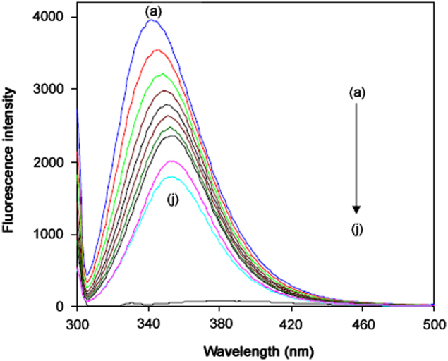
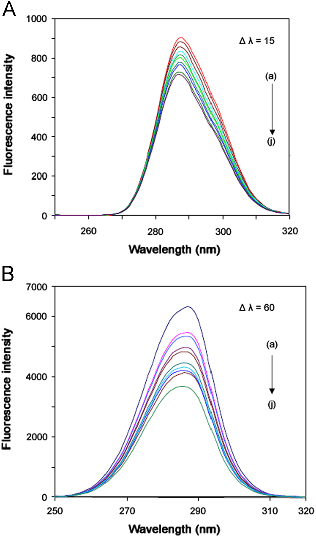
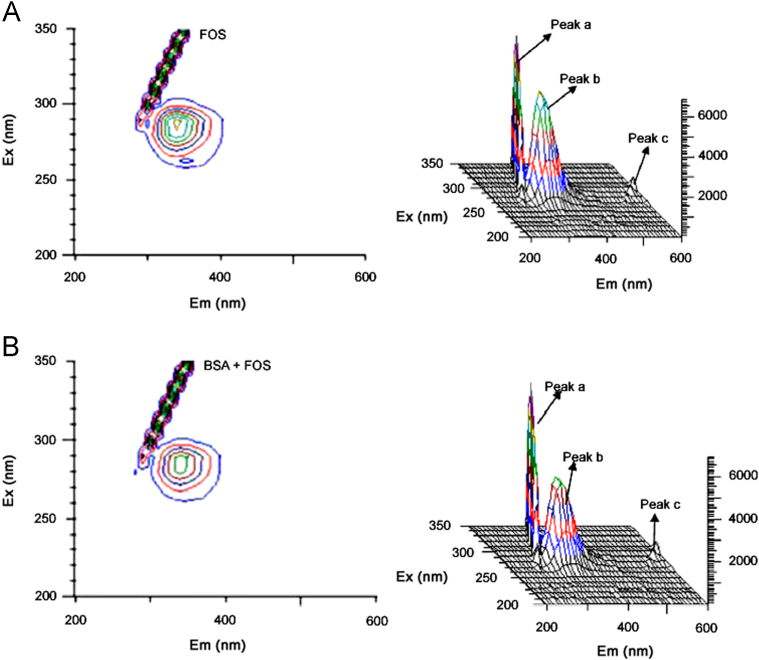
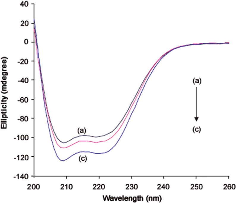
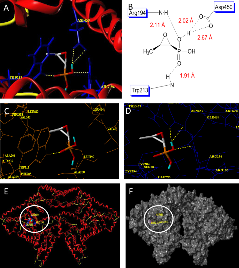
The interaction between fosfomycin (FOS) and bovine serum albumin (BSA) has been investigated effectively by multi-spectroscopic techniques under physiological pH 7.4. FOS quenched the intrinsic fluorescence of BSA via static quenching. The number of binding sites n and observed binding constant KA were measured by the fluorescence quenching method. The thermodynamic parameters ΔG0, ΔH0 and ΔS0 were calculated at different temperatures according to the van't Hoff equation. The site of binding of FOS in the protein was proposed to be Sudlow's site I based on displacement experiments using site markers viz. warfarin, ibuprofen and digitoxin. The distance r between the donor (BSA) and acceptor (FOS) molecules was obtained according to the Förster theory. The effect of FOS on the conformation of BSA was analyzed using synchronous fluorescence spectra (SFS), circular dichroism (CD) and 3D fluorescence spectra. A molecular modeling study further confirmed the binding mode obtained by the experimental studies.
Keywords: 3D spectra; Fosfomycin; Serum albumin; Spectroscopic methods; Synchronous fluorescence.
Figures
References
-
- Yua J., Lib B., Daia P. Molecular simulation of the interaction between novel type rhodanine derivative probe and bovine serum albumin. Spectrochim. Acta A: Mol. Biomol. Spectrosc. 2009;74:277–281. - PubMed
-
- Zhao H.W., Ge M., Zhang Z.X. Spectroscopic studies on the interaction between riboflavin and albumins. Spectrochim. Acta A: Mol. Biomol. Spectrosc. 2006;65:811–817. - PubMed
-
- Hendlin D., Stapley E.O., Jackson M. Phosphonomycin, a new antibiotic produced by strams of streptomyces. Science. 1969;166:122–123. - PubMed
-
- Bergogne-Berezin E. In: Fosfomycin and Derivatives in Antimicrobial Agents. Bryskier A., editor. ASM Press; Washington, DC: 2005. pp. 972–982.
LinkOut - more resources
Full Text Sources
Other Literature Sources
