

Lateral Skull Base Approaches in Pediatric Skull Base Surgery
- PMID: 29404241
- PMCID: PMC5796819
- DOI: 10.1055/s-0038-1624572
Lateral Skull Base Approaches in Pediatric Skull Base Surgery
Abstract
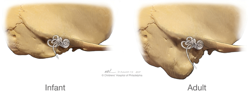
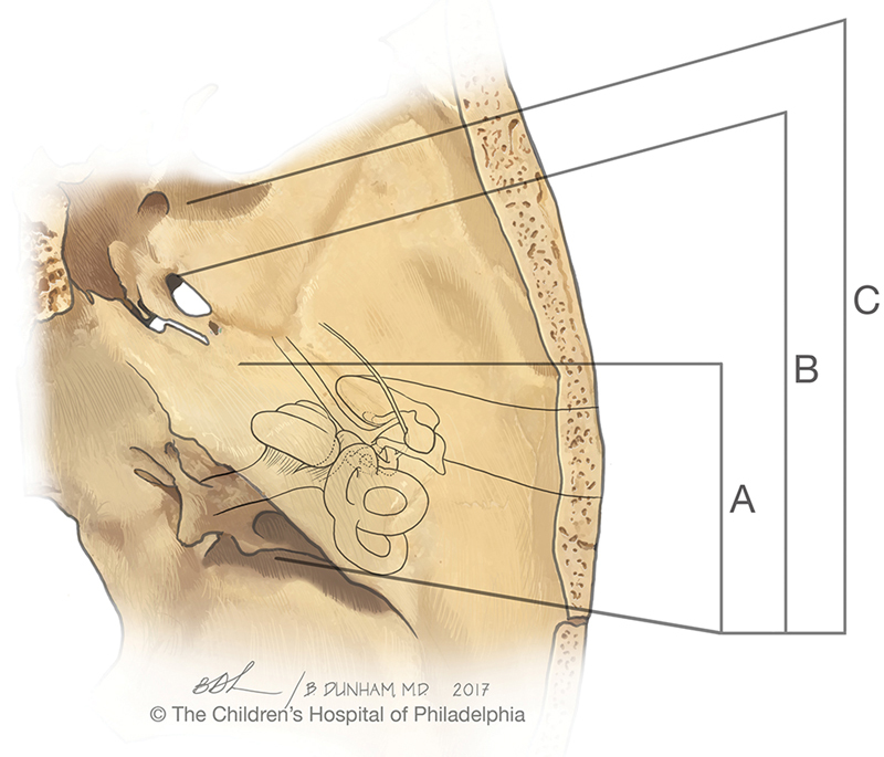
Lateral skull base pathology is rare in children. Awareness of the potential for lateral skull base lesions in children is imperative for timely identification and appropriate management. Some of the common presentations and pathologies shall be presented, as well as a variety of approaches that may be utilized to access the lateral skull base in the pediatric patient. Although the lateral skull base approaches utilized in adults may also be considered for management of pediatric lesions, some special considerations given the small developing anatomy need to be kept in mind.
Keywords: cholesteatoma; endoscopic; lateral skull base; pediatric; pediatric skull base; petrous bone; rhabdomyosarcoma; skull base; surgical approach; temporal bone.
Figures
Similar articles
-
Lateral skull base surgery in a pediatric population: A 25-year experience in a referral skull base center.Int J Pediatr Otorhinolaryngol. 2017 Mar;94:70-75. doi: 10.1016/j.ijporl.2017.01.017. Epub 2017 Jan 14. Int J Pediatr Otorhinolaryngol. 2017. PMID: 28167016
-
Endoscopic transcanal corridors to the lateral skull base: Initial experiences.Laryngoscope. 2015 Sep;125 Suppl 5:S1-13. doi: 10.1002/lary.25203. Epub 2015 Feb 20. Laryngoscope. 2015. PMID: 25703066
-
Developmental Considerations in Pediatric Skull Base Surgery.J Neurol Surg B Skull Base. 2018 Feb;79(1):3-12. doi: 10.1055/s-0037-1617449. Epub 2018 Jan 5. J Neurol Surg B Skull Base. 2018. PMID: 29404235 Free PMC article. Review.
-
Exclusive endoscopic transcanal approach to lateral skull base lesions: Institutional experience of 3 cases.J Otol. 2021 Jan;16(1):55-60. doi: 10.1016/j.joto.2020.08.004. Epub 2020 Sep 4. J Otol. 2021. PMID: 33505451 Free PMC article.
-
Open Approaches to the Anterior Skull Base in Children: Review of the Literature.J Neurol Surg B Skull Base. 2018 Feb;79(1):42-46. doi: 10.1055/s-0037-1621739. Epub 2018 Jan 24. J Neurol Surg B Skull Base. 2018. PMID: 29404240 Free PMC article. Review.
Cited by
-
Pediatric multicompartmental trigeminal schwannoma: illustrative case.J Neurosurg Case Lessons. 2021 May 17;1(20):CASE2171. doi: 10.3171/CASE2171. eCollection 2021 May 17. J Neurosurg Case Lessons. 2021. PMID: 35855022 Free PMC article.
References
-
- Gil Z, Constantini S, Spektor S et al.Skull base approaches in the pediatric population. Head Neck. 2005;27(08):682–689. - PubMed
-
- Gruber D P, Brockmeyer D. Pediatric skull base surgery. 1. Embryology and developmental anatomy. Pediatr Neurosurg. 2003;38(01):2–8. - PubMed
-
- Teo C, Dornhoffer J, Hanna E, Bower C.Application of skull base techniques to pediatric neurosurgery Childs Nerv Syst 199915(2-3):103–109. - PubMed
-
- Cunningham M J, Myers E N, Bluestone C D. Malignant tumors of the head and neck in children: a twenty-year review. Int J Pediatr Otorhinolaryngol. 1987;13(03):279–292. - PubMed
-
- Grinblat G, Prasad S C, Fulcheri A, Laus M, Russo A, Sanna M. Lateral skull base surgery in a pediatric population: a 25-year experience in a referral skull base center. Int J Pediatr Otorhinolaryngol. 2017;94:70–75. - PubMed
Publication types
LinkOut - more resources
Full Text Sources
Other Literature Sources
