Review
. 2018 Jan 19:8:3.
doi: 10.3389/fcimb.2018.00003. eCollection 2018.

The Life Cycle of L. pneumophila: Cellular Differentiation Is Linked to Virulence and Metabolism

Affiliations
Review

The Life Cycle of L. pneumophila: Cellular Differentiation Is Linked to Virulence and Metabolism

Giulia Oliva et al. Front Cell Infect Microbiol. .

Abstract

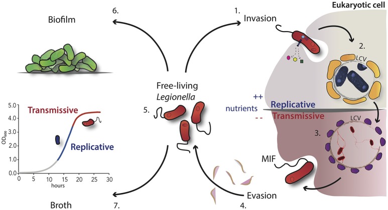
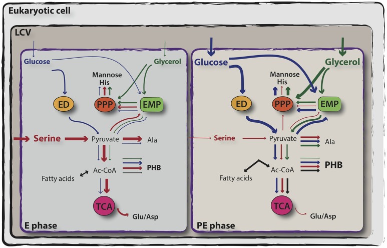
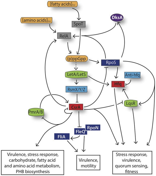
Legionella pneumophila is a gram-negative bacterium that inhabits freshwater ecosystems, where it is present in biofilm or as planktonic form. L. pneumophila is mainly found associated with protozoa, which serve as protection from hostile environments and as replication niche. If inhaled within aerosols, L. pneumophila is also able to infect and replicate in human alveolar macrophages, eventually causing the Legionnaires' disease. The transition between intracellular and extracellular environments triggers a differentiation program in which metabolic as well as morphogenetic changes occur. We here describe the current knowledge on how the different developmental states of this bacterium are regulated, with a particular emphasis on the stringent response activated during the transition from the replicative phase to the infectious phase and the metabolic features going in hand. We propose that the cellular differentiation of this intracellular pathogen is closely associated to key metabolic changes in the bacterium and the host cell, which together have a crucial role in the regulation of L. pneumophila virulence.

Keywords: Legionella pneumophila; life cycle; metabolism; regulation; virulence.


Figures

Figure 1
Figure 1
Schematic overview of the L. pneumophila morphological states during its growth cycle. 1. Uptake of virulent L. pneumophila by the host cell like protozoa or macrophages through convention or coiling phagocytosis. 2. After internalization, the bacteria evade the phagosome-lysosome fusion and start the intracellular multiplication within the LCV, which is surrounded by vesicles (in yellow) rich in lipids and proteins. 3. Nutrient starvation induces the activation of the stringent response and morphological changes. Bacteria express the transmissive traits such as motility (flagella) and become cytotoxic. 4. These infectious bacteria are able to lyse the vacuolar membrane and are released in the extracellular environment. 5. Free-living transmissive bacteria may start a new cycle or persist in the extracellular environment as planktonic form. 6. Alternatively, L. pneumophila may be associated within biofilms, either in natural fresh-water habitats or artificial ones. 7. In broth culture, L. pneumophila displays also a biphasic life cycle, which closely mimics the replicative and transmissive intracellular forms.
Figure 2
Figure 2
Simplified representation of exponential and post-exponential phase-dependent utilization of serine, glucose and glycerol by L. pneumophila. In vitro isotopologue labeling experiments using 13C-serine, 13C-glucose and 13C-glycerol revealed a bipartite metabolism in which serine (in red) is mainly used during the exponential phase of growth and enters primarily the TCA cycle, whereas glucose (in blue) and glycerol (in green) are shuffled into anabolic processes during the post-exponential growth phase of L. pneumophila. Relative carbon fluxes are depicted by the thickness of the arrows. For more detail, see the text. ED, Entner–Doudoroff pathway; PPP, pentose phosphate pathway; EMP, Embden-Meyerhof-Parnas pathway (glycolysis); TCA, tricarboxylic acid cycle (adapted from Eisenreich and Heuner, 2016).
Figure 3
Figure 3
Model the stringent response network governing L. pneumophila differentiation. Amino acid and fatty acid starvation triggers RelA and SpoT to produce the alarmone (p)ppGpp. Its accumulation induces the activation of the stress sigma factor RpoS, the LetA/LetS TCS and consequently an increased transcription of the RsmZ, RsmX, and RsmY sRNAs. The three sRNAs act as “sponge,” sequestering CsrA and leading thereby to the activation of the infectious traits and to changes in the metabolism. The dashed arrows indicate suggested but not yet confirmed direct interactions.

References

    1. Al-Bana B. H., Haddad M. T., Garduño R. A. (2014). Stationary phase and mature infectious forms of Legionella pneumophila produce distinct viable but non-culturable cells. Environ. Microbiol. 16, 382–395. 10.1111/1462-2920.12219 - DOI - PubMed
    1. Albert-Weissenberger C., Cazalet C., Buchrieser C. (2007). Legionella pneumophila- a human pathogen that co-evolved with fresh water protozoa. Cell Mol. Life Sci. 64, 432–448. 10.1007/s00018-006-6391-1 - DOI - PMC - PubMed
    1. Albert-Weissenberger C., Sahr T., Sismeiro O., Hacker J., Heuner K., Buchrieser C. (2010). Control of flagellar gene regulation in Legionella pneumophila and its relation to growth phase. J. Bacteriol. 192, 446–455. 10.1128/JB.00610-09 - DOI - PMC - PubMed
    1. Al-Khodor S., Kalachikov S., Morozova I., Price C. T., Abu Kwaik Y. (2009). The PmrA/PmrB two-component system of Legionella pneumophila is a global regulator required for intracellular replication within macrophages and protozoa. Infect. Immun. 77, 374–386. 10.1128/IAI.01081-08 - DOI - PMC - PubMed
    1. Altman E., Segal G. (2008). The response regulator CpxR directly regulates expression of several Legionella pneumophila icm/dot components as well as new translocated substrates. J. Bacteriol. 90, 1985–1996. 10.1128/JB.01493-07 - DOI - PMC - PubMed

Publication types