Review
. 2018 Jan 19:6:2.
doi: 10.3389/fped.2018.00002. eCollection 2018.

Advances in Diagnosis and Management of Hemodynamic Instability in Neonatal Shock

Affiliations
Review

Advances in Diagnosis and Management of Hemodynamic Instability in Neonatal Shock

Yogen Singh et al. Front Pediatr. .

Abstract

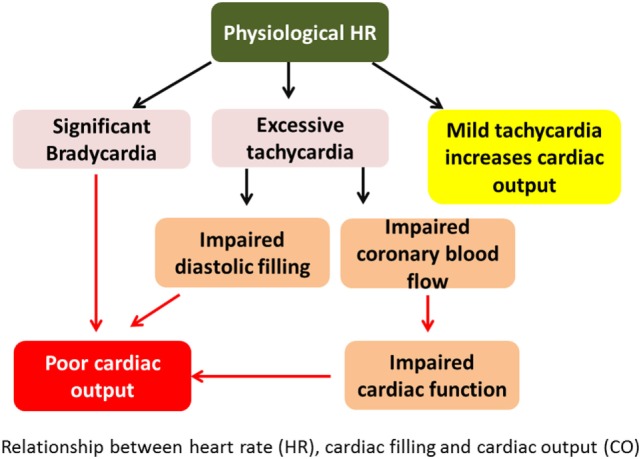
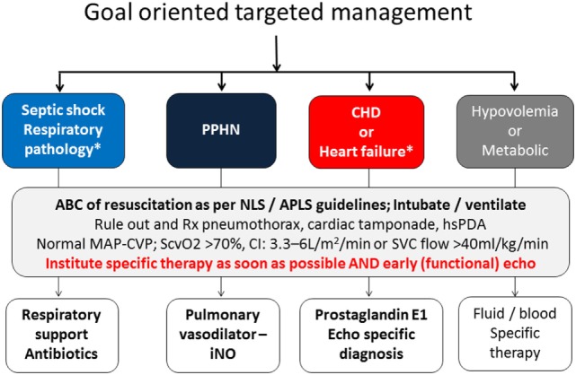
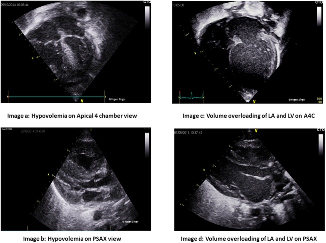
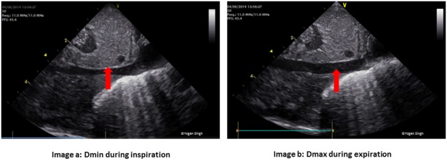
Shock in newborn infants has unique etiopathologic origins that require careful assessment to direct specific interventions. Early diagnosis is key to successful management. Unlike adults and pediatric patients, shock in newborn infants is often recognized in the uncompensated phase by the presence of hypotension, which may be too late. The routine methods of evaluation used in the adult and pediatric population are often invasive and less feasible. We aim to discuss the pathophysiology in shock in newborn infants, including the transitional changes at birth and unique features that contribute to the challenges in early identification. Special emphasis has been placed on bedside focused echocardiography/focused cardiac ultrasound, which can be used as an additional tool for early, neonatologist driven, ongoing evaluation and management. An approach to goal oriented management of shock has been described and how bed side functional echocardiography can help in making a logical choice of intervention (fluid therapy, inotropic therapy or vasopressor therapy) in infants with shock.

Keywords: cardiac output; functional echocardiography; hemodynamic; neonatal shock; tissue perfusion.


Figures

Figure 1
Figure 1
Relationship between heart rate (HR), cardiac filling, and cardiac output (CO). Excessive tachycardia may decrease CO by decreasing preload and hence stroke volume. It may also impair cardiac function from decreased coronary perfusion in shortened diastole.
Figure 2
Figure 2
Goal oriented targeted management and role of echocardiography in instituting specific intervention.
Figure 3
Figure 3
Assessment on cardiac filling on visual inspection “eyeballing.” Images (A,B) show under-filled heart in apical 4 chamber (A4C) and parasternal long axis (PLAX) views. Images (C,D) show volume overloading of left atrium (LA) and left ventricle (LV) in A4C and PLAX views.
Figure 4
Figure 4
Physiological variation in inferior vena cava (IVC) diameter. Normal collapsibility of (A) IVC during inspiration (Dmin) and (B) expansion during expiration (Dmax). In hypovolemia, IVC may be collapsed while in hypervolemia there is minimal or no collapsibility.
Figure 5
Figure 5
Pericardial effusion in (A) subcostal and (B) apical 4 chamber views. In large pericardial effusion and cardiac tamponade, there may be collapse of cardiac chambers—first seen collapse of right atrium followed by right ventricle.
Figure 6
Figure 6
Interventricular septum (IVS) and left ventricle (LV) shape in pulmonary hypertension on visual inspection. Image (A) shows normal circular LV and IVS shapes. Image (B) shows right ventricular dilatation and hypertrophy of right ventricle, flattening of IVS and “D” shaped LV in pulmonary hypertension.
Figure 7
Figure 7
Quantitative assessment of pulmonary artery systolic pressure (PAP) by measuring tricuspid valve regurgitation velocity [tricuspid regurgitation (TR) jet]. PAP = right atrial (RA) pressure + pressure gradient between RA and RV (estimated by TR jet). (A) TR jet on A4C. (B) TR Doppler.

Comment in

Similar articles

Cited by

References

    1. Brierley J, Carcillo JA, Choong K, Cornell T, Decaen A, Deymann A, et al. Clinical practice parameters for hemodynamic support of pediatric and neonatal septic shock: 2007 update from the American College of Critical Care Medicine. Crit Care Med (2009) 37:666–88.10.1097/CCM.0b013e31819323c6 - DOI - PMC - PubMed
    1. Anderson PA. The heart and development. Semin Perinatol (1996) 20:482–509.10.1016/S0146-0005(96)80064-4 - DOI - PubMed
    1. Rowland DG, Gutgesell HP. Noninvasive assessment of myocardial contractility, preload, and afterload in healthy newborn infants. Am J Cardiol (1995) 75:818–21.10.1016/S0002-9149(99)80419-6 - DOI - PubMed
    1. Kluckow M, Evans N. Superior vena cava flow in newborn infants: a novel marker of systemic blood flow. Arch Dis Child Fetal Neonatal Ed (2000) 82:F182–7.10.1136/fn.82.3.F182 - DOI - PMC - PubMed
    1. Davies JM, Tweed WA. The regional distribution and determinants of myocardial blood flow during asphyxia in the fetal lamb. Pediatr Res (1984) 18:764–7.10.1203/00006450-198408000-00019 - DOI - PubMed

LinkOut - more resources