. 2018 Jan 31;4(1):eaap9004.
doi: 10.1126/sciadv.aap9004. eCollection 2018 Jan.

Correction of diverse muscular dystrophy mutations in human engineered heart muscle by single-site genome editing

Affiliations

Correction of diverse muscular dystrophy mutations in human engineered heart muscle by single-site genome editing

Chengzu Long et al. Sci Adv. .

Abstract

Genome editing with CRISPR/Cas9 is a promising new approach for correcting or mitigating disease-causing mutations. Duchenne muscular dystrophy (DMD) is associated with lethal degeneration of cardiac and skeletal muscle caused by more than 3000 different mutations in the X-linked dystrophin gene (DMD). Most of these mutations are clustered in "hotspots." There is a fortuitous correspondence between the eukaryotic splice acceptor and splice donor sequences and the protospacer adjacent motif sequences that govern prokaryotic CRISPR/Cas9 target gene recognition and cleavage. Taking advantage of this correspondence, we screened for optimal guide RNAs capable of introducing insertion/deletion (indel) mutations by nonhomologous end joining that abolish conserved RNA splice sites in 12 exons that potentially allow skipping of the most common mutant or out-of-frame DMD exons within or nearby mutational hotspots. We refer to the correction of DMD mutations by exon skipping as myoediting. In proof-of-concept studies, we performed myoediting in representative induced pluripotent stem cells from multiple patients with large deletions, point mutations, or duplications within the DMD gene and efficiently restored dystrophin protein expression in derivative cardiomyocytes. In three-dimensional engineered heart muscle (EHM), myoediting of DMD mutations restored dystrophin expression and the corresponding mechanical force of contraction. Correcting only a subset of cardiomyocytes (30 to 50%) was sufficient to rescue the mutant EHM phenotype to near-normal control levels. We conclude that abolishing conserved RNA splicing acceptor/donor sites and directing the splicing machinery to skip mutant or out-of-frame exons through myoediting allow correction of the cardiac abnormalities associated with DMD by eliminating the underlying genetic basis of the disease.


Figures

Fig. 1
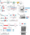
Fig. 1. Myoediting strategy and identification of optimal guide RNAs to target the top 12 exons in DMD.
(A) Conserved splice sites contain multiple NAG and NGG sequences, which enable cleavage by SpCas9. The numbers indicate the frequency of occurrence (%). (B) Human DMD exon structure. Shapes of intron-exon junctions indicate complementarity that maintains the open reading frame upon splicing. Red arrowheads indicate the top 12 targeted exons. The numbers indicate the order of the exons. (C) T7E1 assays in human 293 cells transfected with plasmids expressing the corresponding guide RNA (gRNA), SpCas9, and GFP for the top 12 exons. The PCR products from GFP+ and GFP cells were cut with T7 endonuclease I (T7E1), which is specific to heteroduplex DNA caused by CRISPR/Cas9-mediated genome editing. Red arrowhead indicates cleavage bands of T7E1. bp indicates the base pair length of the marker bands.
Fig. 2
Fig. 2. Rescue of dystrophin mRNA expression in iPSC-derived cardiomyocytes with diverse mutations by myoediting.
(A) Schematic of the myoediting of DMD iPSCs and 3D-EHMs–based functional assay. (B) Myoediting targets the exon 51 splice acceptor site in Del DMD iPSCs. A deletion (exons 48 to 50) in a DMD patient creates a frameshift mutation in exon 51. The red box indicates out-of-frame exon 51 with a stop codon. Destruction of the exon 51 splice acceptor in DMD iPSCs allows splicing from exons 47 to 52 and restoration of the dystrophin open reading frame. (C) Using the guide RNA library, three guide RNAs (Ex51-g1, Ex51-g2, and Ex51-g3) that target sequences 5′ of exon 51 were selected. (D) RT-PCR of cardiomyocytes differentiated from uncorrected DMD (Del), corrected DMD (Del-Cor.), and WT iPSCs. Skipping of exon 51 allows splicing from exons 47 to 52 (lower band) and restoration of the DMD open reading frame. (E) Myoediting strategy for pseudo-exon 47A (pEx). DMD exons are represented as blue boxes. Pseudo-exon 47A (red) with stop codon is marked by a stop sign. The black box indicates myoediting-mediated indel. (F) Sequence of guide RNAs for pseudo-exon 47A of pEx. DMD exons are represented as blue boxes, and pseudo-exons are represented as red boxes (47A). sgRNA, single-guide RNA. (G) RT-PCR of human cardiomyocytes differentiated from WT, uncorrected DMD (pEx), and corrected DMD iPSCs (pEx-Cor.) by guide RNAs In47A-g1 and In47A-g2. Skipping of pseudo-exon 47A allows splicing from exons 47 to 48 (lower band) and restoration of the DMD open reading frame. (H) Myoediting strategy for the duplication (Dup) of exons 55 to 59. DMD exons are represented as blue boxes. Duplicated exons are represented as red boxes. The black box indicates myoediting-mediated indel. (I) Sequence of guide RNAs for intron 54 of Dup (In54-g1, In54-g2, and In54-g3). (J) RT-PCR of human cardiomyocytes differentiated from WT, uncorrected DMD (Dup), and corrected DMD iPSCs (Dup-Cor.). Skipping of duplicated exons 55 to 59 allows splicing from exons 54 to 55 and restoration of the DMD open reading frame. RT-PCR of RNA was performed with the indicated sets of primers (F and R) (table S2).
Fig. 3
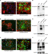
Fig. 3. Immunocytochemistry and Western blot analysis show dystrophin protein expression rescued by myoediting.
(A to C) Immunocytochemistry of dystrophin expression (green) shows DMD iPSC cardiomyocytes lacking dystrophin expression. Following successful myoediting, the corrected DMD iPSC cardiomyocytes express dystrophin. Immunofluorescence (red) detects cardiac marker troponin-I. Nuclei are labeled by Hoechst dye (blue). (D to F) Western blot analysis of WT (100 and 50%), uncorrected (Del, pEx, and Dup) and corrected DMD (Del-Cor#27, pEx-Cor#19, and Dup-Cor#6.) iCM. Red arrowhead (above 250 kD) indicates the immunoreactive bands of dystrophin. Blue arrowhead (above 150 kD) indicates the immunoreactive bands of MyHC loading controls. kD indicates protein molecular weight. Scale bar, 100 μm. Uncropped Western blots with Del-Cor., pEx-Cor., Dup-Cor., and other single colonies are presented in fig. S5.
Fig. 4
Fig. 4. Rescued DMD cardiomyocyte-derived EHM showed enhanced FOC.
(A) Experimental setup for EHM preparation, culture, and analysis of contractile function. (B to D) Contractile dysfunction in DMD EHM can be rescued by myoediting. FOC normalized to muscle content of each individual EHM in response to increasing extracellular calcium concentrations; n = 8/8/6/4/6/6/4/4; *P < 0.05 by two-way analysis of variance (ANOVA) and Tukey’s multiple comparison test. WT EHM data are pooled from parallel experiments with indicated DMD lines and applied to Fig. 4 (B to D). (E) Maximal cardiomyocyte FOC (at 4 mM extracellular calcium) normalized to WT. n = 8/8/6/4/6/6/4/4; *P < 0.05 by one-way ANOVA and Tukey’s multiple comparison test. (F) Titration of corrected cardiomyocytes revealed that 30% of cardiomyocytes needed to be repaired to partially rescue the phenotype, and 50% of cardiomyocytes needed to be repaired to fully rescue the phenotype (100% Del-Cor.) in EHMs. WT, Del, and 100% Del-Cor. are pooled data, as displayed in Fig. 4B.

References

    1. Mendell J. R., Shilling C., Leslie N. D., Flanigan K. M., al-Dahhak R., Gastier-Foster J., Kneile K., Dunn D. M., Duval B., Aoyagi A., Hamil C., Mahmoud M., Roush K., Bird L., Rankin C., Lilly H., Street N., Chandrasekar R., Weiss R. B., Evidence-based path to newborn screening for Duchenne muscular dystrophy. Ann. Neurol. 71, 304–313 (2012). - PubMed
    1. Fairclough R. J., Wood M. J., Davies K. E., Therapy for Duchenne muscular dystrophy: Renewed optimism from genetic approaches. Nat. Rev. Genet. 14, 373–378 (2013). - PubMed
    1. Fayssoil A., Nardi O., Orlikowski D., Annane D., Cardiomyopathy in Duchenne muscular dystrophy: Pathogenesis and therapeutics. Heart Fail. Rev. 15, 103–107 (2010). - PubMed
    1. Jinek M., Chylinski K., Fonfara I., Hauer M., Doudna J. A., Charpentier E., A programmable dual-RNA-guided DNA endonuclease in adaptive bacterial immunity. Science 337, 816–821 (2012). - PMC - PubMed
    1. Cong L., Ran F. A., Cox D., Lin S., Barretto R., Habib N., Hsu P. D., Wu X., Jiang W., Marraffini L. A., Zhang F., Multiplex genome engineering using CRISPR/Cas systems. Science 339, 819–823 (2013). - PMC - PubMed

Publication types