. 2018 Apr;31(2):201-209.
doi: 10.1007/s10278-018-0055-1.

A General Framework for Monitoring Image Acquisition Workflow in the Radiology Environment: Timeliness for Acute Stroke CT Imaging

Affiliations

A General Framework for Monitoring Image Acquisition Workflow in the Radiology Environment: Timeliness for Acute Stroke CT Imaging

Timothy P Szczykutowicz et al. J Digit Imaging. 2018 Apr.

Abstract

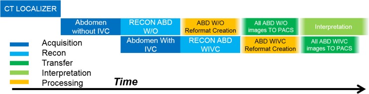
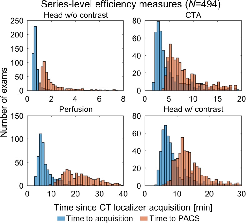
Many facets of an image acquisition workflow leave a digital footprint, making workflow analysis amenable to an informatics-based solution. This paper describes a detailed framework for analyzing workflow and uses acute stroke response timeliness in CT as a practical demonstration. We review methods for accessing the digital footprints resulting from common technologist/device interactions. This overview lays a foundation for obtaining data for workflow analysis. We demonstrate the method by analyzing CT imaging efficiency in the setting of acute stroke. We successfully used digital footprints of CT technologists to analyze their workflow. We presented an overview of other digital footprints including but not limited to contrast administration, patient positioning, billing, reformat creation, and scheduling. A framework for analyzing image acquisition workflow was presented. This framework is transferable to any modality, as the key steps of image acquisition, image reconstruction, image post processing, and image transfer to PACS are common to any imaging modality in diagnostic radiology.

Keywords: CT; Compliance; Informatics; Quality control; Radiology workflow; Stroke.


Conflict of interest statement

All authors are involved in a collaborative project that supplies CT protocols to GE Healthcare. TPS is also a GE consultant and the founder of protocolshare.org. RB is co-founder and officer of ImageMoverMD, and GW is on the MAB of McKesson and HealthMyne, a stockholder in HealthMyne, and co-founder of WITS(MD). AF receives research support from GE Healthcare. WP is a stockholder in GE Healthcare.

Figures

Fig. 1
Fig. 1
Overview of the temporal relationship between the major steps involved with imaging exam workflow. Time progresses as one moves from left to right. Boxes overlapping in the vertical direction represent workflow tasks that may occur at the same point in time
Fig. 2
Fig. 2
Overview of the temporal relationship between the major steps involved with just the imaging acquisition, reconstruction and post processing, and networking facets of diagnostic medical imaging workflow. This example is for a two phase abdomen exam with axial reconstructions and reformats for both the with and without IVC contrast series. Time progresses as one moves from left to right. Boxes overlapping in the vertical direction represent workflow tasks that may occur at the same point in time. The order, time between, and duration of the events in this figure all provide temporal efficiency information on both the operator and scanner/hardware/software
Fig. 3
Fig. 3
Representative histograms of institution-level analysis. Histograms indicate distributions of time to series acquisition (blue) and availability in PACS (orange) in 494 patients over approximately 1.5 years. All PACS times refer to the first recon (not reformats) other than for perfusion, which refers to perfusion maps. All times are referenced to the time of CT localizer radiograph acquisition

References

    1. Boland GW. Enhancing CT productivity: strategies for increasing capacity. American Journal of Roentgenology. 2008;191(1):3–10. doi: 10.2214/AJR.07.3208. - DOI - PubMed
    1. Gunn ML, Maki JH, Hall C, Bhargava P, Andre JB, Carnell J, ... Beauchamp NJ: Improving MRI scanner utilization using modality log files. J Am Coll Radiol 14(6):783–786, 2017. - PubMed
    1. Flug J, Nagy P. The lean concept of waste in radiology. Journal of the American College of Radiology. 2011;8(6):443–445. doi: 10.1016/j.jacr.2011.02.017. - DOI - PubMed
    1. Hirschorn DS, Hinrichs CR, Gor DM, Shah K, Visvikis G. Impact of a diagnostic workstation on workflow in the emergency department at a level I trauma center. Journal of digital imaging. 2001;14:199–201. doi: 10.1007/BF03190338. - DOI - PMC - PubMed
    1. Reiner BI, Siegel EL, Hooper FJ, Pomerantz S, Dahlke A, Rallis D. Radiologists’ productivity in the interpretation of CT scans: a comparison of PACS with conventional film. American Journal of Roentgenology. 2001;176(4):861–864. doi: 10.2214/ajr.176.4.1760861. - DOI - PubMed

Publication types

MeSH terms